Transformative Potential of Nonmetal doped Transition Metal Oxide Nanoparticles for Multifaceted Applications
1University Ddepartment of Chemistry Ranchi University Ranchi Jharkhand, India.
2Department of Agronomy, Institute of Agricultural Sciences, BHU, Varanasi, Uttar Pradesh, India.
Corresponding Author E-mail:dr.smriti.singh@gmail.com
DOI : http://dx.doi.org/10.13005/msri/220107
Download this article as:
![]()
Nonmetal doped transition metal oxide nanoparticles (TMONPs) have attracted much attention from researchers due to their novel magnetic, optical, and electrochemical properties. Due to such distinct features of TMONPs, these are deemed suitable in applications such as super-capacitors, lithium-ion batteries, non-volatile memory circuits, energy management devices, energy storage, and conversion applications such as solar cells, and infrared detectors. From these discoveries outlined, the identification of specific developments such as catalysis, energy storage, environmental remediation, and biomedical applications can be outdone. This chapter represents a critical evaluation of the synthesis methods, doping techniques, characterization, and recent applications of non-metal doped TMONPs.
KEYWORDS:Biomedical nanotechnology; Electrocatalysis Environmental remediation; Nonmetal doping; Photocatalysis; Transition Metal Oxides
Introduction
Overview of Transition Metal Oxides
Transition metal oxides (TMOs) are compounds formed by oxygen atoms bonded to transition metals, elements found in groups 3-12 of the modern periodic table. These oxides tend to have diverse characteristics in terms of physical properties like high melting point, conductivity, variable oxidation states, etc., and chemical properties due to the diverse electronic configurations. Of the TMOs identified in Table 1, both the dopant and host materials exhibit a diverse range of applications, including catalytic, electronic, optical, and magnetic applications specific to primarily titanium dioxide (TiO₂), zinc oxide (ZnO), and iron oxide (Fe₂O₃)1 . Figure 1 showcases the latest advancements in Transition Metal Oxides.
The chemical behaviour of TMOs is a result of the d-electrons being responsible for different oxidation states of the transition metals2 and thus forming different bonds. It also allows TMOs to display low dielectric constants, flexibility in the band gap, and magnetic structures. For this reason, they are employed in various applications ranging from photo-catalysis for environmental remediation, energy storage in lithium-ion batteries, 3 and optically transparent conductive oxides for optoelectronics devices4 .
 |
Figure 1: Recent advances in Transition Metal Oxides. |
Advantages and Potential Applications of Nonmetals-Doped TMO nanoparticles (TMONPs)
Doping involves the introduction of impurities into a metal or semiconductor to deliberately alter its electrical, thermal, or structural properties. Further, doping can enhance conductivity, modify band structure, and improve the overall performance of the material for specific applications. Doping with nonmetal anions or molecules also shows a significant effect on the TMONPs by affecting the electronic structure. The chemical state and location of the dopant are crucial to the effectiveness of the doping process. The purpose of doping transition metal oxides with nonmetals is to enhance their intrinsic properties like optical properties, thermal stability, electrical conductivity, magnetic properties, etc., and expand their applications. This chapter focuses on the recent progress in the nonmetal doping of TMONPs 5.
Nonmetal dopants, such as nitrogen, sulphur, and carbon, lead to new energy levels within the band structure of the TMOs, thereby changing their electrical, optical, and catalytic properties6. For instance, doping of nitrogen into TiO₂ can considerably decrease the band gap,7 which enables the material to be more effective in photocatalytic applications. Besides, doping enhances the electrochemical properties of supercapacitors by providing higher energy density. Likewise, when sulphur is doped into ZnO, it can also show improved photocatalytic and photoelectrochemical behaviour because the presence of defective anions facilitates charge separation, thereby reducing the rate of recombination8. Also, sulphur-doped TiO₂ can improve lithium-ion diffusion9, leading to higher battery capacity and faster charging.
Zhang et al10. proposed that Carbon-doping into ZnO nanoparticles resulted in the reduced bandgap of ZnO, enhanced the separation efficiency of electron-hole pairs, and significantly boosted the photocatalytic performance under visible light.
Doping can also improve the charge carrier mobility, which is essential for applications in electronics and photovoltaics. Kim et al11. reported that Nitrogen-doped TiO₂ has been keen on exhibiting higher electron mobility, resulting in better performance in dye-sensitized solar cells.
Nonmetal doping also participates in the regulation of the surface chemistry of TMONPs, which is crucial for catalytic processes12 .Through the alteration of surface properties, nonmetal doped TMONPs serve more active sites for the reaction to happen and hence improve the catalytic activity. This mainly benefits photocatalytic water splitting, CO₂ reduction, and pollutant degradation reactions.
Further, doping in TMONPs provides biomedical advantages such as biocompatibility, targeted drug delivery, and good contrast for imaging. For instance, carbon-doped ZnO nanoparticles can serve as potential photodynamic agents for therapeutic applications and bioimaging13. Figure 2 shows the potential applications of nonmetal doped TMONPs.
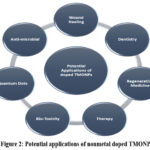 |
Figure 2: Potential applications of nonmetal doped TMONPs. |
Furthermore, doping with nonmetals can develop the thermal and chemical stability of TMONPs, making them stronger for industrial applications. Incorporating nonmetals can also facilitate the synthesis of nanostructured TMOs with controlled morphology and size, further enhancing their application potential [Refer to Table 1].
Table 1: Some examples of TMONPs with nonmetal dopant and their applications.
| NON-METAL DOPANT | TMONPs | SYNTHESIS PROCEDURE | APPLICATIONS | REFERENCES |
| Nitrogen (N) | TiO2 | conventional precipitation route | photocatalytic hydrogen generation | [69-91] |
| Sol-gel method | Photocatalysis | |||
| Solvothermal synthesis | Degradation of benzene | |||
| Hydrothermal Synthesis | dye-sensitization of solar cells | |||
| ZnO | Hydrothermal Synthesis | Photocatalysis | ||
| Sol-gel method | Visible photocatalytic degradation | |||
| Fe2O3 | Direct in-situ synthesis | photo-electrochemical properties | ||
| Sulfur (S) | TiO2 | Scaffold template technique | photocatalytic dye degradtion | |
| Ball Milling Process | Dielectric Properties | |||
| Hydrothermal synthesis | Degradation of methyl orange | |||
| ZnO | Sol-gel Method | Antibacterial activities | ||
| hydrothermal method | Optical properties study | |||
| α‐ Fe2O3 | Microwave synthesis | Photodegradation of methyl orange | ||
| Carbon (C) | TiO2 | Sol-gel | Visible-light photocatalysis | |
| Hydrothermal synthesis | Use in optical devices | |||
| ZnO | Hydrothermal synthesis | Photocatalytic performance | ||
| Sol-gel | Magnetic properties | |||
| Phosphorous (P) | Co₃O₄ | Hydrothermal Method | Hydrogen evolution | |
| TiO2 | Sol-gel Method | Photocatalytic properties under the solar light irradiation | ||
| ZnO | Sol-gel Method | Structural and Optical Properties study | ||
| Iodine (I) | TiO2 | Sol-gel Method | Photocatalytic and antibacterial activities | |
| Hydrothermal synthesis | Improved dye degradation. |
Purpose and Scope of the Chapter
The present chapter aims to provide a comprehensive overview to disseminate the current understanding of the TMONPs doped with nonmetals.
It aims at providing the methods used in doping and the changes that have occurred in the structural, optical, electrical, and magnetic properties, due to nonmetal doping.
The areas highlighted for focus include the identification of several synthesis techniques.
It also discusses various characterization methods used to describe the structural efficiency and functionality of doped TMONPs.
In addition, the chapter focuses on their versatile applications in the field the energy and environment.
Materials and Methods
Synthesis of Transition Metal Oxide Nanoparticles
Sol-gel Method
TMONPs can be synthesized using the sol-gel method, which entails transitioning a liquid “sol” (mostly colloidal) into a solid “gel” phase. For instance, titanium dioxide (TiO₂) nanoparticles can be synthesized from titanium alkoxides precursor, which on hydrolysis and poly-condensation produces a gel that on further drying and calcination, yields nanoparticles14. MAM Khan et al15. studied the synthesis of ZnO nanoparticles with different Mn doping concentrations by sol-gel wet chemical route. The sol-gel technique provides good control over the composition and structure of the nanoparticles, making it suitable for applications like catalysis and photo-catalysis 16.
This method involves converting a liquid solution (“sol”) into a solid (“gel”) phase. For example, titanium dioxide (TiO₂) nanoparticles can be made using titanium alkoxides. The process includes following steps
Hydrolysis: Titanium alkoxides react with water.
Condensation: The products of hydrolysis react to form a gel.
Gel Formation: A three-dimensional network forms, creating a gel.
Drying and Calcination: The gel is dried and heated to produce nanoparticles.
Hydrothermal Synthesis
Hydrothermal synthesis involves the process of forming nanoparticles under high temperature and high-pressure conditions using water as a solvent, which is crucial for a well-crystallized and homogeneous structure of nanoparticles. Mohan et al. employed a hydrothermal method for the synthesis of zinc oxide (ZnO) nanoparticles using zinc nitrate and sodium hydroxide17. The hydrothermal method is preferable when it concerns synthesizing nanoparticles of high purity that are perfect for use in sensors, electronics, and photocatalytic applications 18,19,20.
This method uses high temperature and pressure with water to form well-crystallized nanoparticles.
Dissolution: Metal precursors dissolve in water.
Nucleation: High temperature and pressure cause nanoparticles to form.
Crystal Growth: Temperature and time control the size of particles.
Post-Treatment: The product is washed and dried.
Co-precipitation Method
Co-precipitation synthesis method involves the instantaneous precipitation of multiple components from a homogeneous solution. For example, Bolivar et al. prepared iron oxide (Fe₃O₄) nanoparticles with a mixture of ferric and ferrous ions that are precipitated with a base such as ammonia, followed by magnetic field precipitation 21. Vijayaprasath et al. synthesized Ni/Mn co-doped ZnO nanoparticles to study their structural and magnetic behavior22. Gandhi et al. synthesized cobalt doped ZnO nanoparticles by co-precipitation method and studied their magnetic properties 23. This method is particularly useful for the mass production of nanoparticles bearing uniform size and composition, suitable for magnetic storage and bioimaging 24.
Here, multiple components precipitate from a solution at the same time.
Ionic Mixing: Metal ions mix in a solution.
Precipitation: A base is added to form insoluble hydroxides.
Aging and Transformation: The hydroxides change into metal oxides.
Separation and Drying: The product is filtered, washed, and dried.
Other relevant methods
Other synthesis processes comprise combustion synthesis25, microwave-assisted synthesis26, and electrochemical deposition 27.
Combustion Synthesis
Precursor Preparation: Mix metal precursors with a fuel.
Ignition: Trigger an exothermic reaction to generate heat.
Particle Formation: Particles form during combustion.
Cooling and Collection: Collect particles after cooling.
Microwave-Assisted Synthesis
Precursor Solution: Dissolve metal salts in a solvent.
Microwave Heating: Apply uniform heating to promote rapid particle formation.
Particle Growth: Control reaction conditions to shape and size particles.
Cooling and Collection: Collect and purify the nanoparticles.
Electrochemical Deposition
Electrolysis: Reduce metal ions at the cathode.
Deposition: Regulate voltage to control film thickness.
Crystallization: Improve nanoparticle quality through heating.
Michalow et al 28. employed the combustion method to synthesize Niobium-doped titanium dioxide nanoparticles by burning a pre-formed paste made of a titanium precursor and a niobium salt.
Falk et.al29 synthesized anatase TiO2 nanoparticles by microwave-assisted synthesis to induce high photocatalytic activity with increased crystallinity and phase transformation . Electrochemical deposition offers the opportunity to fine-tune both the size and composition of the deposited nanoparticles using the electrochemical parameters. Altogether, these synthesis methods offer flexible routes to synthesize high-performance TMONPs.
Summary of Benefits, Challenges, and Efficiency
Combustion Synthesis
Rapid, high-yield process with a simple setup but prone to particle agglomeration. Efficient for large-scale production but offers limited control over particle size and morphology.
Microwave-Assisted Synthesis
Fast, energy-efficient method with uniform heating but requires specialized equipment. Highly efficient but limited to materials that absorb microwave radiation.
Electrochemical Deposition
Provides precise control over film thickness and composition but has a complex setup. Efficient for thin films but restricted to conductive substrates.
Figure 3 shows a few other methods to synthesize nonmetal doped TMONPs.
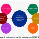 |
Figure 3: Methods to synthesize nonmetal doped TMONPs. |
Doping Techniques
Chemical Doping
Chemical doping involves introducing dopant atoms into the host material during synthesis by evenly distributing dopants within the host material. For instance, Darzi et al. synthesized nitrogen-doped titanium dioxide (TiO₂) nanoparticles using a sol-gel method with an acid catalysis process, resulting in the desired narrowing of the band gap and an enhancement in the photocatalytic activity 30. Than et al. synthesized N-doped TiO2 nanoparticles, where the nitrogen atoms replace oxygen atoms in the TiO₂ lattice, resulting in a reduced band gap and enhanced visible-light-driven photocatalytic activity 31.
Physical Doping
Physical doping refers to techniques where dopant atoms are physically introduced into the host nanoparticles, often after the initial synthesis. This can be done through ball milling, thermal diffusion, or sputtering32. Cho et al. successfully synthesized sulfur-doped ZnO nanostars using a chemical vapor deposition method 33 and studied the significant influence of sulfur doping on the morphology of ZnO nanostructures, paving the way for photocatalysis, sensing, and other fields.
Ion implantation
Ion implantation is the process where the dopant ions are purposely directed and embedded in the host material with the help of an ion beam 34. As reported by Shieh et al., cobalt ions can indeed affect the optical properties of TiO₂ thin films leading to enhanced visible light response and different photoluminescence characteristics 35.
The doping techniques mentioned above and many other methods such as Spin-coating36, In-situ doping 37, Photolithography38, and Diffusion doping are some of the most efficient ways of increasing the properties of transition metal oxide nanoparticles, enabling their use in a broad range of applications. Table 2 shows the TMONPs with nonmetal doping and their key properties.
Table 2: Nonmetal doped TMONPs with their key properties
| TMONPs | Doping Element | Synthesis Method | Key Properties | References |
| TiO2 | N | Precipitation Method | Reduced band gap, enhanced visible light absorption | 65-69 |
| Fe2O3 | C | Sol-gel method | Enhanced magnetic properties, Increased surface area | |
| ZnO | S | Combustion method | Improved electrical conductivity, Additional active sites | |
| NiO | B | Pyrolysis method | Improved thermal stability, Enhanced electrochemical performance | |
| Co3O4 | N | Hyderthermal Method | Enhanced catalytic activity, reduced activation energy |
Characterization Techniques
Table 3: Showing Characterization techniques and its significance
| Sl.No. | Characterization Method | Description | Purpose | Key Parameter Analyzed | Significance | Alternate Techniques |
| 1 | UV-Vis Spectroscopy | Measures absorbance across UV-visible wavelengths. | Analyze optical properties of nanoparticles | Band gap energy, absorbance spectrum |
Provides size and electronic structure information. |
Photolumin escence Spectroscopy (PL), Raman Spectroscopy |
| 2 | FTIR (Fourier Transform Infrared Spectroscopy) |
Measures infrared absorption to analyze functional groups. | Identify chemical bonds and functional groups | Vibrational frequencies of functional groups |
Identifies chemical bonds and surface functiona lization. |
Raman Spectroscopy, XPS (X-ray Photoelectron Spectroscopy) |
| 3 | XRD (X-ray Diffraction) |
Analyzes crystal structure and phase purity. | Determine crystalline structure | Crystal structure, phase identification | Provides crystallinity and phase identification. |
SAED (Selected Area Electron Diffraction), TEM |
| 4 | FESEM (Field Emission Scanning Electron Microscopy) |
Captures high-resolution images for surface morphology. | Visualize surface morphology | Particle size, surface topography |
Determines nanoparticle morphology and size distribution. | TEM, AFM (Atomic Force Microscopy) |
| 5 | EDX (Energy Dispersive X-ray Spectroscopy) |
Performs elemental analysis by detecting X-ray emissions. | Identify elemental composition | Elemental composition, impurity detection | Provides elemental composition and distribution. | ICP-MS (Inductively Coupled Plasma Mass Spectrometry), XPS |
| 6 | Zeta Potential |
Measures surface charge and stability in colloidal suspensions. | Measure colloidal stability | Surface charge, stability | Provides insight into colloidal stability and aggregation. | DLS (Dynamic Light Scattering), Electrophoretic Mobility |
| 7 | Impedance Spectroscopy | Measures electrical impedance across frequencies. | Analyze electrical properties | Conductivity, resistivity, dielectric constant | Provides electrical and dielectric properties. |
Electrochemical Impedance Spectroscopy (EIS), Conductivity Measurements |
| 8 | BET Surface Area Analysis |
Measures surface area via gas adsorption. | Measure surface area and porosity | Surface area, pore volume |
Provides surface area, pore volume, and distribution, crucial for catalysis. |
BJH (Barrett- Joyner- Halenda) Method, Mercury Porosimetry |
| 9 | TGA/DTA (Thermo gravimetric Analysis/ Differential Thermal Analysis) |
Measures weight loss and heat flow with temperature variation. | Analyze thermal stability | Weight loss, decomposi tion tempera ture |
Provides thermal stability and decomposi tion information. |
DSC (Differential Scanning Calorimetry) |
| 10 | NMR (Nuclear Magnetic Resonance) |
C13 and proton NMR analyze molecular structure and dynamics. | Determine molecular structure | Chemical environ ment, bonding |
Provides chemical environment and structure information. | Mass Spectrometry (MS), EPR (Electron Paramagnetic Resonance) |
| 11 | Cyclic Voltammetry | Studies redox behavior via cyclic voltage sweeps. | Analyze redox properties | Redox activity, electron transfer | Provides electrochemical properties and reaction kinetics. | Potentiostatic Methods, Chronoam perometry |
X-ray Diffraction (XRD)
X-ray diffraction (XRD) is extensively used to identify phases, assess the crystalline quality, and determine the crystallite size in transition metal oxide-based nanoparticles. Doping affects the crystal structure by disrupting the crystalline symmetry of the material, altering the lattice constants, inhibiting structural distortions, and reducing crystallinity 39. So, crystallographic analysis can also reveal how doping affects the crystal structure to enhance material properties 40.
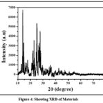 |
Figure 4: Showing XRD of Materials. |
Scanning Electron Microscopy (SEM)
SEM provides surface images of nanoparticles illustrating their morphology and size. Nair et al. explained that the SEM images of undoped ZnO have a slightly smaller grain size than Co-doped ZnO 41. Vu et al. illustrated the morphology of nonmetal doped TiO2 nanotubes with a diameter ranging from 20 to 30 nm and a length of 150 to 200 nm 42.
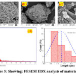 |
Figure 5: Showing: FESEM EDX analysis of materials |
Transmission Electron Microscopy (TEM)
TEM offers high-resolution imaging and can identify the lattice structure of the nanoparticles. Anandan et al. showed the particle diameter estimation via TEM, which revealed an average diameter of approximately 29 nm for pure TiO2 and 21 nm for 1 wt% IO3– doped TiO2 43. Ahmmad et al 44. analyzed the TEM images and explained that each sub-microsphere comprises aggregated TiO2 nanoparticles ranging in size from 20 to 40 nm, exhibiting rough surfaces, confirming the polycrystalline nature of the anatase and rutile phases within the TiO2 sub-microspheres.
Fourier-Transform Infrared (FTIR) and Raman Spectroscopy
FTIR and Raman spectroscopy can identify chemical bonds and molecular structures, essential for analyzing dopant effects on the nanoparticle. Khalid et al. studied the FTIR spectra for GO, TiO₂, and GR-TiO₂ composite samples, revealing the disappearance of the C=O peak at 1720 cm⁻¹ in the GR-TiO₂ sample, which signifies the reduction of GO to graphene during the hydrothermal process45. Caratto et al46. prepared Nitrogen-doped TiO2 nanoparticles on glass substrates to verify homogeneity. Raman spectroscopy analysis reveals a peak at 146 cm⁻¹, related to O–Ti–O bending vibrations, shifted towards lower wavenumbers (up to 140 cm⁻¹ for 15% ammonia), indicating the presence of N3- ions in interstitial sites.
Other Techniques
Several other techniques are also gaining popularity for characterizing transition metal oxide nanomaterials. These techniques include UV-visible spectroscopy 47, Atomic Force Microscopy (AFM) 48, Brunauer-Emmett-Teller (BET) analysis 49, Thermogravimetric Analysis (TGA) 50, and Differential Thermal Analysis (DTA). Researchers also utilize TGA/DTA to study the thermal performance of samples. Energy-dispersive X-ray (EDX) 51, often coupled with SEM or TEM, provides elemental analysis and mapping, confirming the presence and distribution of dopants.
Zeta Potentials
The zeta potential is a measure of the surface effective electric charge of nanoparticles, which is determined by their charge stability. The zeta potential indicates the particle’s ability to move when an electric field is applied. The stability of particles improves with increasing magnitude, with particles agglomerating in the 0-5 mV range, remaining somewhat stable in the 20-40 mV range, and being highly stable at 40 mV and above. The zeta potential was determined using the Zetasizer Nano ZS.
Table 4: Showing Zeta potential value and its significance
| Application | Description | Zeta Potential Range (mV) | Significance of Material (26.2 mV) |
| Water Treatment | Positive zeta potential attracts and absorbs negatively charged contaminants (e.g., anionic dyes, organic pollutants, heavy metal anions). | +20 to +40 (moderate stability) | The material’s zeta potential (+26.2 mV) ensures sufficient stability for coagulation and adsorption in water purification. |
| Suitable for coagulation and flocculation processes in water purification. | |||
| Catalysis | Positive surface charge enhances interactions with negatively charged reactants, facilitating specific oxidation or reduction reactions. | +20 to +30 | The material’s charge allows it to interact effectively with anionic reactants, benefiting catalytic applications. |
| Drug Delivery | Positive zeta potential aids in interacting with negatively charged cell membranes for targeted drug delivery. | +20 to +30 (stable dispersion) | The material’s moderate stability ensures good dispersion in biological fluids while retaining its charge for drug delivery. |
| Moderate stability ensures dispersion in biological fluids while allowing aggregation under specific conditions for controlled drug release. | |||
| Agriculture | Positively charged particles interact with negatively charged soil components or plant roots, aiding in the delivery of nutrients or agrochemicals. | +20 to +40 (suitable stability) | The material’s stability and positive charge make it effective for delivering nutrients or agrochemicals in soils. |
| Energy Storage | Materials with moderate stability and uniform charge distribution are suitable for supercapacitors or batteries. | +20 to +40 | The material’s zeta potential ensures uniform dispersion in liquid electrolytes, enhancing its potential as an electrode. |
| Positive zeta potential suggests potential use as an electrode material due to good dispersion in liquid electrolytes. | |||
| Nanomaterials in Coatings or Inks | Monodispersity and positive charge ensure even distribution, making the material ideal for uniform film formation in coatings. | +20 to +40 | The material’s positive charge and stability enhance its application in coatings, ensuring uniform distribution. |
| It can be used in stable ink or paint formulations. |
Results
The study examined the effects of nonmetal doping on the properties of transition metal oxide nanoparticles (TMONPs). Results showed that XRD analysis confirmed changes in lattice parameters, with nitrogen-doped TiO₂ showing reduced crystallite size, promoting better surface reactivity. SEM and TEM revealed uniform, smaller particles with increased surface roughness, crucial for catalytic and electrochemical applications. UV-Vis spectroscopy showed a bandgap reduction in nitrogen and carbon-doped ZnO, enhancing visible light absorption. FTIR spectra confirmed dopant-induced vibrational shifts, highlighting modifications in chemical bonds. Impedance spectroscopy showed increased conductivity in sulfur and nitrogen-doped ZnO, attributed to improved charge carrier mobility and reduced recombination rates. Cyclic voltammetry revealed enhanced redox activity, particularly in sulfur-doped NiCoOx nanorods. Nonmetal-doped TMONPs demonstrated higher photocatalytic degradation rates of organic pollutants, while carbon-doped ZnO showed superior phenol degradation under visible light.
Discussions
Nonmetal-doped TMONPs exhibit enhanced performance due to synergistic effects between the host lattice and dopants. Dopants create lattice distortions, forming oxygen vacancies and mid-gap states, which reduce crystallite size and increase active surface area. Carbon and sulfur doping introduce mid-gap states near the conduction band, enabling visible light absorption and broadening the material’s applicability in photocatalysis and solar cells. Sulfur doping introduces defective anions, facilitating charge separation, leading to increased photocatalytic efficiency and electrochemical performance. Nitrogen doping enhances electron mobility, benefiting supercapacitors and dye-sensitized solar cells. Surface chemistry changes due to nonmetal doping create more reactive sites, making these materials suitable for pollutant degradation, hydrogen evolution reactions, and CO₂ reduction. Carbon-doped ZnO’s photodynamic properties and stability support targeted drug delivery and bioimaging, while Ag-Fe₂O₃ nanoparticles show potential as antibacterial agents. Nonmetal doping enhances thermal and chemical stability, crucial for industrial scalability, and maintains structural integrity under high-temperature applications, supporting their use in supercapacitors and energy devices.
Applications of Nonmetal Doped TMONPs
Photocatalysis
Nonmetal doped TMONPs exhibit enhanced photocatalytic properties due to their unique electrical properties, high surface area, suitable band gap, and presence of active sites 52. Due to these properties, they are widely used in various photocatalytic applications, such as photocatalytic water splitting including both oxygen evolution reaction (OER) and hydrogen evolution reaction (HER). Ansari et al53. reported that N-doped TiO₂ enhances photocatalytic efficiency enabling broad solar spectrum absorption for water splitting reactions. The nitrogen doping subsequently reduces the band gap enabling it to absorb visible light and generate more charge carriers to facilitate the splitting of water molecules to produce oxygen and hydrogen, contributing to sustainable hydrogen production for fuel cells. Zhang et al 54. studied that Sulfur doped ZnO facilitated oxidation of CO and reduction of NOx, contributing to air purification and emission control . Qu et al 55. demonstrated the potential of phosphorus-doped cobalt sulfides as efficient and stable electro-catalysts for hydrogen evolution, highlighting the importance of rational design in developing advanced catalytic materials. Figure 4 shows the advantages of nonmetal doped TMNOPs for photocatalytic applications.
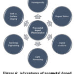 |
Figure 6: Advantages of nonmetal doped TMNOPs as photocatalysts. |
Electrocatalysis
Nonmetal doped TMONPs serve as efficient electrocatalysts to accelerate electrochemical reactions essential for energy conversion and storage technologies. Li et al56. stated that S-doped NiCoOx nanorods exhibit efficient OER catalysis with increased active surface area and enhanced electronic states due to sulfur incorporation, further improving water-splitting performance, and highlighting a promising system for practical hydrogen production.
Pollutant Degradation
Nonmetal doped TMONPs play an active role in the degradation of pollutants in both water and air purification processes. Yu et al. demonstrated that C-TiO₂ nanoparticles exhibit superior visible light photocatalytic activity for phenol degradation due to effective charge transfer and prolonged lifetime of photogenerated charges 57. Similarly, N-doped TiO₂ nanoparticles have demonstrated effective degradation of airborne pollutants under visible light by facilitating the breakdown of various air pollutants as studied by Wang et al. 58
Supercapacitors
Supercapacitors bridge the gap between batteries and dielectric capacitors by offering both higher power density and higher energy density. The electrode materials used in supercapacitors need to be conductive and possess a high surface area 59. Consequently, TMONPs, are favored because of their superior specific capacitance and extended cycle life 60. Lin et al. showed that nitrogen-doped carbon nanotubes (CNTs) combined with transition metal oxides like MnO₂ NPs result in supercapacitors with high specific capacitance with excellent cyclic stability 61. Vinothkumar et al62. synthesized calcium-doped copper oxide nanoparticles to be used as an efficient bifunctional electrocatalyst for various supercapacitor applications.
Drug Delivery and Bioimaging
Nonmetal doping increases the surface area and active sites of TMONPs, allowing for higher drug loading capacity. It can improve the responsiveness of TMONPs to external stimuli (e.g., pH, temperature), enabling controlled and sustained release of therapeutic agents.
El-Bassuony et al64. reported Ag-Fe₂O₃ nanoparticles as potential antibacterial agents in drug delivery systems and for magnetic resonance imaging (MRI) contrast enhancement [63]. Bajpai et al. investigated the synthesis and multifunctional applications of nitrogen and phosphorus-doped carbon nanodots (N, P-doped CNDs). The research highlights the utility of N, P-doped CNDs in cellular bio-imaging. Their strong fluorescence properties enable effective imaging of cells, making them suitable for diagnostic and research purposes .
Table 5: A comparative analysis of non-metal doped and undoped transition metal oxide nanoparticles
| Property | Undoped Transition Metal Oxide Nanoparticles | Non-Metal Doped Transition Metal Oxide Nanoparticles |
| Band Gap Energy | Typically wide band gaps, limiting visible light absorption. | Reduced band gaps, enhancing visible light absorption. For instance, sulfur-doped ZnO exhibits a band gap of 2.81 eV, compared to 3.2 eV for undoped ZnO. |
| Photocatalytic Activity | Limited activity under visible light due to larger band gaps. | Enhanced activity under visible light. Sulfur-doped ZnO showed an 81.4% degradation efficiency for organic dyes under solar irradiation. |
| Structural Properties | Retain intrinsic crystal structures with minimal defects. | Introduction of dopants can induce defects or oxygen vacancies, influencing properties. For example, nitrogen-doped ZnO nanoparticles exhibit a reduced crystallite size of 12 nm. |
| Mechanism of Property Enhancement | Properties are intrinsic to the material’s composition. | Dopants introduce mid-gap states or modify electronic structures, facilitating visible light absorption and improved charge separation. Nitrogen doping in ZnO creates localized states above the valence band, reducing the band gap. |
Conclusion
The doping of nonmetal into transition metal oxides showcases highly improved structural, optical, electrical, and magnetic properties of the TMONPs. It is feasible to use sol-gel, hydrothermal, or co-precipitation synthesis methods to produce doped TMONPs. The characterization techniques which include XRD, SEM, TEM, and EDX, can assist in determining the structural and morphological parameters. Nonmetal doping of TMONPs finds applications in catalysis, energy storage, environmental, and biomedical sectors, which reveals their multifunctional and versatile applications. Nonmetal doped TMONPs hold significance in advancing both environmental remediation and energy applications. Their potential to degrade industrial pollutants in ambient conditions will lead to advancements in photocatalytic environmental remediation. Further, nonmetal doped TMONPs play an important role in sustainable energy through their superior electrical and optical properties, thereby increasing the efficiency of solar cells, fuel cells, and supercapacitors.
Ongoing research is also concentrating on innovative doping technologies, including plasma-assisted doping and atomic layer deposition, to gain precise oversight on dopant distribution and concentration for improved performance. Interdisciplinary techniques combining materials science, chemistry, and engineering are being applied to fully realize the potential of nonmetal doped TMONPs to resolve some global issues related to environment and energy contributing to a cleaner and sustainable world.
Acknowledgment
We would like to acknowledge the University Department of Chemistry Ranchi University , Ranchi, Jharkhand for their kind support and for providing technical support. The authors are thankful to the Department of Agronomy, Institute of Agricultural Science BHU for their kind support.
Funding Sources
The author(s) have not received any financial support for this study, authorship, publication of this article.
Conflict of Interest
The authors do not have any conflict of interest.
Data Availability Statement
This statement does not apply to this article.
Ethics Statement
This research did not involve human participants, animal subjects, or any material that requires ethical approval.
Informed Consent Statement
This study did not involve human participants, and therefore, informed consent was not required.
Permission to reproduce material from other sources
Not applicable
Author contributions
Rajesh Kumar: Conceptualization, writing original draft and data interpretation.
Shruti Jaiswal: Designed the chapter content and data analysis.
Rudhima Raj: Draft and data analysis.
Dr. Anil Kumar Delta: supervision, writing original draft and data interpretation.
Dr. Smriti Singh: Conceptualization, visualization, supervision, writing original draft and data interpretation
References
- Kumar RR, Kumar KU, Haranath D. Synthesis, properties, and applications of transition metal oxide nanomaterials. In: Multifunctional Nanostructured Metal Oxides for Energy Harvesting and Storage Devices. CRC Press; 2020:1-73.
CrossRef - Akbari A, Amini M, Tarassoli A, et al. Transition metal oxide nanoparticles as efficient catalysts in oxidation reactions. Nano-Structures and Nano-Objects. 2018;14:19-48.
CrossRef - Wu HB, Chen JS, Hng HH, Lou XWD. Nanostructured metal oxide-based materials as advanced anodes for lithium-ion batteries. Nanoscale. 2012;4(8):2526-2542.
CrossRef - Diao F, Wang Y. Transition metal oxide nanostructures: premeditated fabrication and applications in electronic and photonic devices. J Mater Sci. 2018;53:4334-4359.
CrossRef - Yadav P, Dwivedi PK, Tonda S, et al. Metal and nonmetal doped metal oxides and sulfides. In: Green Photocatalysts. 2020:89-132.
CrossRef - Al-Naggar AH, Shinde NM, Kim JS, Mane RS. Water splitting performance of metal and non-metal-doped transition metal oxide electrocatalysts. Coord Chem Rev. 2023;474:214864.
CrossRef - Morikawa T, Asahi R, Ohwaki T, Aoki K, Taga Y. Band-gap narrowing of titanium dioxide by nitrogen doping. Jpn J Appl Phys. 2001;40(6A):L561.
CrossRef - Mirzaeifard Z, Shariatinia Z, Jourshabani M, Rezaei Darvishi SM. ZnO photocatalyst revisited: effective photocatalytic degradation of emerging contaminants using S-doped ZnO nanoparticles under visible light radiation. Ind Eng Chem Res. 2020;59(36):15894-15911.
CrossRef - Zhang Y, He X, Tang J, et al. Sulfur-doped TiO2 anchored on a large-area carbon sheet as a high-performance anode for sodium-ion battery. ACS Appl Mater Interfaces. 2019;11(47):44170-44178.
CrossRef - Zhang X, Qin J, Hao R, et al. Carbon-doped ZnO nanostructures: facile synthesis and visible light photocatalytic applications. J Phys Chem C. 2015;119(35):20544-20554.
CrossRef - Kim SB, Park JY, Kim CS, et al. Effects of graphene in dye-sensitized solar cells based on nitrogen-doped TiO2 composite. J Phys Chem C. 2015;119(29):16552-16559.
CrossRef - Marschall R, Wang L. Nonmetal doping of transition metal oxides for visible-light photocatalysis. Catal Today. 2014;225:111-135.
CrossRef - Mishra DK, Mohapatra J, Sharma MK, et al. Carbon doped ZnO: Synthesis, characterization and interpretation. J Magn Magn Mater. 2013;329:146-152.
CrossRef - Sungur Ş. Titanium dioxide nanoparticles. In: Handbook of Nanomaterials and Nanocomposites for Energy and Environmental Applications. 2021:713-730.
CrossRef - Khan MM, Kumar S, Alhazaa AN, Al-Gawati MA. Modifications in structural, morphological, optical and photocatalytic properties of ZnO: Mn nanoparticles by sol-gel protocol. Mater Sci Semicond Process. 2018;87:134-141.
CrossRef - Peña-Garcia R, Guerra Y, Farias BVM, et al. Unusual thermal dependence of saturation magnetization in zinc oxide nanoparticles doped with transition metals obtained by sol gel method. Ceram Int. 2019;45(1):918-929.
CrossRef - Mohan S, Vellakkat M, Aravind A, Reka U. Hydrothermal synthesis and characterization of Zinc Oxide nanoparticles of various shapes under different reaction conditions. Nano Express. 2020;1(3):030028.
CrossRef - Preethi S, Anitha A, Arulmozhi M. A comparative analysis of the properties of zinc oxide (ZnO) nanoparticles synthesized by hydrothermal and sol-gel methods. Indian J Sci Technol. 2016.
CrossRef - Nandagudi A, Nagarajarao SH, Santosh MS, et al. Hydrothermal synthesis of transition metal oxides, transition metal oxide/carbonaceous material nanocomposites for supercapacitor applications. Mater Today Sustain. 2022;19:100214.
CrossRef - Ge S, Shi X, Sun K, et al. Facile hydrothermal synthesis of iron oxide nanoparticles with tunable magnetic properties. J Phys Chem C. 2009;113(31):13593-13599.
CrossRef - Marimón-Bolívar W, González EE. Green synthesis with enhanced magnetization and life cycle assessment of Fe3O4 nanoparticles. Environ Nanotechnol Monit Manag. 2018;9:58-66.
CrossRef - Vijayaprasath G, Murugan R, Asaithambi S, et al. Structural and magnetic behavior of Ni/Mn co-doped ZnO nanoparticles prepared by co-precipitation method. Ceram Int. 2016;42(2):2836-2845.
CrossRef - Gandhi V, Ganesan R, Abdulrahman Syedahamed HH, Thaiyan M. Effect of cobalt doping on structural, optical, and magnetic properties of ZnO nanoparticles synthesized by coprecipitation method. J Phys Chem C. 2014;118(18):9715-9725.
CrossRef - Farahmandjou M, Soflaee F. Synthesis and characterization of α-Fe2O3 nanoparticles by simple co-precipitation method. Phys Chem Res. 2015;3(3):191-196.
- Li FT, Ran J, Jaroniec M, Qiao SZ. Solution combustion synthesis of metal oxide nanomaterials for energy storage and conversion. Nanoscale. 2015;7(42):17590-17610.
CrossRef - Bhatt AS, Bhat DK, Tai CW, Santosh MS. Microwave-assisted synthesis and magnetic studies of cobalt oxide nanoparticles. Mater Chem Phys. 2011;125(3):347-350.
CrossRef - Maduraiveeran G, Sasidharan M, Jin W. Earth-abundant transition metal and metal oxide nanomaterials: Synthesis and electrochemical applications. Prog Mater Sci. 2019;106:100574.
CrossRef - Michalow KA, Flak D, Heel A, et al. Effect of Nb doping on structural, optical and photocatalytic properties of flame-made TiO2 nanopowder. Environ Sci Pollut Res. 2012;19:3696-3708.
CrossRef - Falk GS, Borlaf M, López-Muñoz MJ, Fariñas JC, Rodrigues Neto JB, Moreno R. Microwave-assisted synthesis of TiO2 nanoparticles: photocatalytic activity of powders and thin films. J Nanopart Res. 2018;20:1-10.
CrossRef - Darzi SJ, Mahjoub AR, Sarfi S. Visible-light-active nitrogen doped TiO2 nanoparticles prepared by sol–gel acid catalyzed reaction. Iran J Mater Sci Eng. 2012;9(3):17-23.
- Than LD, Luong NS, Ngo VD, et al. Highly visible light activity of nitrogen doped TiO2 prepared by sol–gel approach. J Electron Mater. 2017;46:158-166.
CrossRef - Muğlu GM. Synthesis of transition-metal-oxide-based nanomaterials by sputtering. In: The Trends In Nano Materials Synthesis and Applications. 2022;137.
- Cho J, Lin Q, Yang S, et al. Sulfur-doped zinc oxide (ZnO) Nanostars: Synthesis and simulation of growth mechanism. Nano Res. 2012;5:20-26.
CrossRef - Yildirim O, Cornelius S, Smekhova A, et al. The local environment of cobalt in amorphous, polycrystalline and epitaxial anatase TiO2: Co films produced by cobalt ion implantation. J Appl Phys. 2015;117(18).
CrossRef - Shieh YN, Chang YY. Influence of cobalt ion implantation on optical properties of titanium dioxide thin films. Thin Solid Films. 2010;518(24):7464-7467.
CrossRef - Vijayaprasath G, Murugan R, Ravi G, et al. Characterization of dilute magnetic semiconducting transition metal doped ZnO thin films by sol–gel spin coating method. Appl Surf Sci. 2014;313:870-876.
CrossRef - Elmehasseb I, Kandil S, Elgendy K. Advanced visible-light applications utilizing modified Zn-doped TiO2 nanoparticles via non-metal in situ dual doping for wastewater detoxification. Optik. 2020;213:164654.
CrossRef - Xia F, Shao Z, He Y, et al. Surface charge transfer doping via transition metal oxides for efficient p-type doping of II–VI nanostructures. ACS Nano. 2016;10(11):10283-10293.
CrossRef - Pu S, Xue S, Yang Z, et al. In situ co-precipitation preparation of a superparamagnetic graphene oxide/Fe3O4 nanocomposite as an adsorbent for wastewater purification: synthesis, characterization, kinetics, and isotherm studies. Environ Sci Pollut Res. 2018;25:17310-17320.
CrossRef - Lo Presti L, Ceotto M, Spadavecchia F, et al. Role of the nitrogen source in determining structure and morphology of n-doped nanocrystalline TiO2. J Phys Chem C. 2014;118(9):4797-4807.
CrossRef - Nair MG, Nirmala M, Rekha K, Anukaliani A. Structural, optical, photo catalytic and antibacterial activity of ZnO and Co doped ZnO nanoparticles. Mater Lett. 2011;65(12):1797-1800.
CrossRef - Vu TA, Dao CD, Hoang TT, et al. Study on photocatalytic activity of TiO2 and non–metal doped TiO2 in Rhodamine B degradation under visible light irradiation. Int J Nanotechnol. 2013;10(3-4):235-246.
CrossRef - Anandan S, Kathiravan K, Murugesan V, Ikuma Y. Anionic (IO3-) non-metal doped TiO2 nanoparticles for the photocatalytic degradation of hazardous pollutant in water. Catal Commun. 2009;10(6):1014-1019.
CrossRef - Ahmmad B, Kusumoto Y, Islam MS. One-step and large scale synthesis of non-metal doped TiO2 submicrospheres and their photocatalytic activity. Adv Powder Technol. 2010;21(3):292-297.
CrossRef - Khalid NR, Ahmed E, Hong Z, Zhang Y, Ahmad M. Nitrogen doped TiO2 nanoparticles decorated on graphene sheets for photocatalysis applications. Curr Appl Phys. 2012;12(6):1485-1492.
CrossRef - Caratto V, Setti L, Campodonico S, et al. Synthesis and characterization of nitrogen-doped TiO2 nanoparticles prepared by sol–gel method. J Sol-Gel Sci Technol. 2012;63:16-22.
CrossRef - Lam SM, Sin JC, Abdullah AZ, Mohamed AR. Transition metal oxide loaded ZnO nanorods: preparation, characterization and their UV–vis photocatalytic activities. Sep Purif Technol. 2014;132:378-387.
CrossRef - Wang MC, Lin HJ, Wang CH, Wu HC. Effects of annealing temperature on the photocatalytic activity of N-doped TiO2 thin films. Ceram Int. 2012;38(1):195-200.
CrossRef - Saien J, Mesgari Z. Highly efficient visible-light photocatalyst of nitrogen-doped TiO2 nanoparticles sensitized by hematoporphyrin. J Mol Catal A Chem. 2016;414:108-115.
CrossRef - Zhao Y, Qiu X, Burda C. The effects of sintering on the photocatalytic activity of N-doped TiO2 nanoparticles. Chem Mater. 2008;20(8):2629-2636.
CrossRef - Atchudan R, Edison TNJI, Perumal S, Vinodh R, Lee YR. In-situ green synthesis of nitrogen-doped carbon dots for bioimaging and TiO2 nanoparticles@ nitrogen-doped carbon composite for photocatalytic degradation of organic pollutants. J Alloys Compd. 2018;766:12-24.
CrossRef - Yue K, Liu S, Yan S, et al. Analysis of the reaction mechanism of N/S co-doped carbon-based catalysts for low-temperature NH3-SCR reduction of NOx. Sep Purif Technol. 2024;344:127302.
CrossRef - Ansari SA, Khan MM, Ansari MO, Cho MH. Nitrogen-doped titanium dioxide (N-doped TiO2) for visible light photocatalysis. New J Chem. 2016;40(4):3000-3009.
CrossRef - Zhang H, Shi W, Gao N, et al. Highly sensitive and selective gas-phase ethanolamine sensor by doping sulfur into nanostructured ZnO. Sens Actuators B Chem. 2019;296:126633.
CrossRef - Qu G, Wu T, Yu Y, et al. Rational design of phosphorus-doped cobalt sulfides electrocatalysts for hydrogen evolution. Nano Res. 2019;12:2960-2965.
CrossRef - Li C, Wong CH, Lam FLY, Hu X. Highly efficient and robust sulfur-doped nickel-cobalt oxide towards oxygen evolution reaction. Mol Catal. 2020;496:111175.
CrossRef - Yu S, Yun HJ, Kim YH, Yi J. Carbon-doped TiO2 nanoparticles wrapped with nanographene as a high performance photocatalyst for phenol degradation under visible light irradiation. Appl Catal B Environ. 2014;144:893-899.
CrossRef - Wang S, Ding H, Zhao Y, Li Y, Wang W. Fabrication of protective textile with N-doped TiO2 embedded citral microcapsule coating and its air purification properties. Fibers Polym. 2020;21:334-342.
CrossRef - Singhal R, Chaudhary M, Tyagi S, et al. Recent developments in transition metal-based nanomaterials for supercapacitor applications. J Mater Res. 2022;37(13):2124-2149.
CrossRef - Deka S. Nanostructured mixed transition metal oxide spinels for supercapacitor applications. Dalton Trans. 2023;52(4):839-856.
CrossRef - Lin TT, Lai WH, Lü QF, Yu Y. Porous nitrogen-doped graphene/carbon nanotubes composite with an enhanced supercapacitor performance. Electrochim Acta. 2015;178:517-524.
CrossRef - Vinothkumar V, Prasad GV, Chen SM, et al. One-step synthesis of calcium-doped copper oxide nanoparticles as an efficient bifunctional electrocatalyst for sensor and supercapacitor applications. J Energy Storage. 2023;59:106415.
CrossRef - El-Bassuony AA, Abdelsalam HK. Attractive study of the physical properties of silver iron oxide nanoparticles for biomedical applications. Phys Scr. 2023;98(5):055919.
CrossRef - Bajpai VK, Khan I, Shukla S, et al. N, P-doped carbon nanodots for food-matrix decontamination, anticancer potential, and cellular bio-imaging applications. J Biomed Nanotechnol. 2020;16(3):283-303.
CrossRef
Abbreviations Used in the Document:
TMONPs – Transition Metal Oxide Nanoparticles
TMOs – Transition Metal Oxides
TiO₂ – Titanium Dioxide
ZnO – Zinc Oxide
Fe₂O₃ – Iron Oxide
OER – Oxygen Evolution Reaction
HER – Hydrogen Evolution Reaction
SEM – Scanning Electron Microscopy
TEM – Transmission Electron Microscopy
XRD – X-Ray Diffraction
EDX – Energy Dispersive X-Ray Spectroscopy
FTIR – Fourier-Transform Infrared Spectroscopy
BET – Brunauer-Emmett-Teller (Analysis)
TGA – Thermogravimetric Analysis
DTA – Differential Thermal Analysis
MRI – Magnetic Resonance Imaging
CNTs – Carbon Nanotubes
ALD – Atomic Layer Deposition
GO – Graphene Oxide
GR-TiO₂ – Graphene-Titanium Dioxide Composite
CNDs – Carbon Nanodots
Accepted on: 13 March 2025
Second Review by: Dr. Nitin Waghmode
Final Approval by: Dr. Shouxun Ji


![]()
![]()
![]()
