Facile Synthesis and Comprehensive Characterisation of Nanoparticles of Fe2O3 and Further Study of Their Antimicrobial and Antimalarial Efficacy
 , Nutan Sadgir*and Sunil Dhonnar
 Department of Chemistry, Mahatma Gandhi Vidya mandir’ Loknete Vyankatrao Hiray Arts, Science, and Commerce College, Panchavati, Nashik, India
Corresponding author, Email address - nutansadgir@gmail.com
Download this article as:
ABSTRACT:Due to their unique physicochemical properties, iron oxide nanoparticles have become an interesting class of materials that can be used in various technological processes. This study highlights the production of hematite nanoparticles (Fe2O3) by the simple co-precipitation method using primary reactants that are iron(III) chloride hexahydrate (FeCl3 6H2O) and ammonia solution. These nanomaterials were characterized by X-ray diffraction (XRD), Fourier-transform infrared spectroscopy (FTIR), and scanning electron microscopy (SEM). The XRD analysis showed that the formation of crystalline Fe2O3 phase was successful and it had crystallite dimensions calculated as about 40 nm. FTIR analysis showed typical Fe-O bands vibrationally, which justified the creation of pure iron oxide structures. SEM micrographs exhibited the morphology of spherical particles and were made to behave as clusters at high temperatures of calcination. Also, the synthesized Fe2O3 nanoparticles possessed significant antimicrobial and antifungal efficacy against specific pathogenic strains, and this should suggest their future use in biomedical and environmental remediation processes.
KEYWORDS:Antimalarial; Antimicrobial; Fe2O3 nanoparticles; SEM; XRD
Introduction
The field of nanotechnology has emerged as a transformative discipline with substantial implications across multiple scientific domains, including environmental science, medicine, and advanced materials development.1–3 Nanoscale materials exhibit enhanced performance characteristics compared to their bulk counterparts, primarily attributed to their reduced dimensionality and resulting unique physicochemical properties.4–6 Contemporary research has particularly focused on applications in targeted drug delivery systems, biosensing platforms, diagnostic imaging, and heterogeneous catalysis support materials.6–8
Magnetic nanoparticles represent a specialized subset of nanomaterials that demonstrate exceptional magnetic behavior at the nanoscale, distinguishing them from conventional magnetic materials.9 The manipulation of matter at the atomic and molecular level, typically within the sub-100 nanometer range, has enabled the development of materials with unprecedented optical, magnetic, electronic, and catalytic characteristics.10,11 These distinctive properties render nanomaterials invaluable for applications in environmental restoration, energy conversion technologies, biomedical interventions, and electronic devices.12
Iron oxide nanoparticles have garnered considerable attention due to their inherent advantages, including natural abundance, economic feasibility, chemical inertness, biocompatibility, and magnetic responsiveness.11,13 The polymorphic nature of iron oxides encompasses several crystalline phases, including magnetite (Fe₃O₄), maghemite (γ-Fe₂O₃), and hematite (α-Fe₂O₃)14 [8]. Hematite represents the thermodynamically most stable configuration under standard atmospheric conditions and exhibits environmental compatibility.15 As an n-type semiconductor material with a bandgap energy of approximately 2.1 eV, hematite demonstrates suitability for photocatalytic applications, energy storage systems, photoelectrochemical processes, and sensor technologies.16
Contemporary investigations have demonstrated the antimicrobial and antifungal efficacy of iron oxide nanoparticles, which is attributed to their capacity for generating reactive oxygen species (ROS), disrupting membranes, and interfering with metabolism.17 This functionality has stimulated interest in biomedical applications, including wound care products, pharmaceutical delivery vehicles, and antimicrobial surface treatments.18,19 However, the biological activity of nanoparticles demonstrates strong dependence on morphological parameters, surface characteristics, and crystalline structure.20
Multiple synthetic approaches have been developed for iron oxide nanoparticle production, encompassing sol-gel processes, hydrothermal synthesis, solvothermal methods, microemulsion techniques, and thermal decomposition procedures.21 Although these methodologies can produce high-quality nanomaterials, they frequently necessitate elevated temperatures, extended reaction periods, costly precursor materials, or sophisticated instrumentation, thereby limiting industrial scalability and economic viability.22
The co-precipitation method offers significant advantages as a simple, cost-effective, and environmentally benign synthetic route that operates under mild conditions while maintaining excellent control over particle characteristics23. This approach facilitates the production of uniform nanoparticles with tunable properties through parameter optimization 24. The present study aims to synthesize Fe₂O₃ nanoparticles via co-precipitation methodology and conduct comprehensive characterization to evaluate their structural, morphological, and biological properties.
Materials and Methods
Materials
Iron(III) chloride hexahydrate (FeCl₃·6H₂O, 99% purity) was obtained at Sigma Aldrich.The ammonia ( NH3, 25% w/w) solution was purchased at Merck. All experimental procedures were performed with distilled water. The chemicals were taken as received without purification.
Synthesis of Fe₂O₃ Nanoparticles
A modified co-precipitation method was used to prepare Fe2O3 nanoparticles25–27. Iron(III) chloride hexahydrate (5.4 g) was used in this procedure: 100 mL of distilled water was added to the entire mass of the compound, which was under consistent magnetic stirring at room temperature and was aimed at complete dissolution and homogeneity. The iron salt solution was then added slowly but vigorously in the presence of ammonia solution to achieve a pH of about 9-10. The instantaneous formation of a reddish-brown precipitate indicated the appearance of iron oxide25,26. The mixture was stirred and kept at 60oC until further precipitation and growth of particles occurred, which took 2 more hours.
The resulting product was centrifuged at 4000 rpm for 15 minutes, and then washed repeatedly with distilled water and ethanol to remove any remained ions and unreacted species. The dried product was left in an oven at 80oC. Lastly, the dried product was then subjected to 400oC temperature during 3 hours under the air to produce crystalline nanoparticle of Fe2O3.
The given method of synthesis is consistent with known co-precipitation procedures, with pH, temperature, and calcination parameters being vital in avoiding particle size, crystallinity, and phase purity25–27 . The procedure outlined is simple, reproducible and can be applied to the yield of Fe2O3 nanoparticles with the desired characteristics in different applications.
Characterization Techniques
X-ray diffraction (XRD) was used to examine the crystalline structure and phase purity of the synthesized nanoparticles using a Rigaku MiniFlex 600 diffractometer with Cu Ka radiation (λ = 1.5406 Å) and operating at 40 kV and 15 mA. The XRD pattern was captured in the 2 θ range (10-80°) and scan rate of 2°/min.
Fourier-transform infrared (FTIR) spectroscopy was done using a PerkinElmer Spectrum 100 spectrometer in a wavenumber range of 400-4000 cm-1 to identify functional groups and chemical bonding using KBr pellet technique.
Scanning electron microscopy (SEM) was used to analyze the morphology of the surface, as well as particle size distribution on a JEOL JSM-7600F field emission microscope at 15 kV acceleration voltage. Gold sputering of samples was performed to improve the conductivity of samples before SEM analysis.
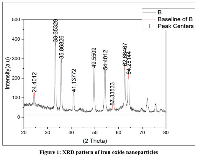 |
Figure 1: XRD pattern of iron oxide nanoparticles Click here to View Figure |
Antimicrobial Activity
Antimicrobialeffect of the synthesized compounds was tested by using the broth microdilution method as per the guidelines of determining minimum inhibitory concentrations (MICs)28,29 11017. The test compounds were produced as stock solutions in DMSO and finally showcased into the variety of broth-media to discount the variety of concentrations of interest in primary and secondary screening. To ascertain solubility of the compounds, the standard bacterial strains were inoculated in each of the wells using DMSO as the diluent to guarantee the presence of the soluble compounds29,30.
In the first screening, the compounds were tested with the concentration of 500, 250, and 200 μg/mL. Active compounds on this stage were further tested on further dilution (100, 62.5, 50, 40, 32, 25 and 12.5 ug/ml) in order to obtain accurate MIC values. Positive controls (bacteria in the absence of antimicrobial agents) and negative controls (media only) were used in each of the assays to confirm the results28–30. Inoculation of the microplates took place followed by incubation at 37C for 1624 hours. Defining of MIC was done in such a way that the least concentration of it did not actually show any growth of microbes at a particular point of time28–30.
Every test was conducted in a trio or at least in duplicate to provide good reproducibility. Control organisms with known susceptibility were used to test the accuracy of the drug concentrations28,30. This standard method helps to compare the antimicrobial efficacy with fair guarantees in a variety of compounds and strains of bacteria.
Results
X-ray Diffraction Analysis
XRD analysis of the synthesized material revealed the formation of pure hematite (α-Fe₂O₃) phase without detectable impurities
FTIR Spectroscopy
FTIR analysis confirmed the formation of Fe₂O₃ nanoparticles through the identification of characteristic vibrational modes.
 |
Figure 2: FTIR of Fe2O3 nanoparticles Click here to View Figure |
Antimicrobial Activity
The broth microdilution method was the method used to determine the antibacterial activity of the synthesized Fe2O3 nanoparticles, on the basis of the methods employed in our earlier study coumarin31,32. The stock solutions of the nanoparticles were prepared through DMSO. The minimum inhibitory concentration values against E. coli (MTCC 443), P. aeruginosa (MTCC 1688), S. aureus (MTCC 96) and S. pyogenus (MTCC 442) were determined.
Discussion
X-ray Diffraction Analysis
XRD analysis of the synthesized material revealed the formation of pure hematite (α-Fe₂O₃) phase without detectable impurities. The diffraction pattern exhibited characteristic peaks at 2θ values of 24.1°, 33.2°, 35.6°, 40.9°, 49.5°, 54.1°, 57.6°, 62.4°, and 64.0°, corresponding to the (012), (104), (110), (113), (024), (116), (018), (214), and (300) crystal planes, respectively. These peaks match well with the standard JCPDS card No. 33-0664 for hematite.
The average crystallite size was calculated using the Debye-Scherrer equation:
D = Kλ/(βcosθ)
where D is the crystallite size, K is the shape factor (0.9), λ is the X-ray wavelength (1.5406 Å), β is the full-width at half maximum (FWHM), and θ is the Bragg angle. The calculated average crystallite size was approximately 40 nm, indicating the successful synthesis of nanocrystalline hematite.
FTIR Spectroscopy
The Fourier transform infrared (FTIR) spectrum of the synthesized Fe2O3 nanoparticles recorded in the region of the frequency range of 4000 to 800 cm-1 is shown in Fig. X. The broad absorption band suggested in the region of 3400-3200 cm-1 is attributed to the O-H stretching vibration of surface hydroxyl and physically adsorbed water molecules. The weak band around 2371 cm-1 is associated with the CO2 in the atmosphere. The bands at around 2178 cm⁻1 and around 2109 cm⁻1 could be combination or overtone vibrations from residual precursor species. The absorption bands appearing at about 1565 cm-1 and at about 1542 cm-1 are attributed to the bending vibration of the -OH groups or the traces left of nitrate residues from the synthesis process. The band at ~1157 cm per cent is attributed to Fe-O-H bending vibration. The characteristic Fe-O stretching vibrations of iron oxide nanoparticles are generally found below 700 cm-1, thus confirming the formation of Fe2O3 nanoparticles. The characteristic Fe–O stretching vibrations below 600 cm⁻¹ could not be clearly observed due to instrumental limitations.
Morphological Analysis
SEM examination revealed that the synthesized Fe₂O₃ nanoparticles exhibited predominantly spherical morphology with relatively uniform size distribution. The particles demonstrated a tendency to form small clusters, which can be attributed to magnetic interactions between individual nanoparticles. The average particle size observed from SEM images was consistent with the crystallite size calculated from XRD analysis. At higher calcination temperatures, increased agglomeration was observed, likely due to enhanced particle mobility and sintering effects.
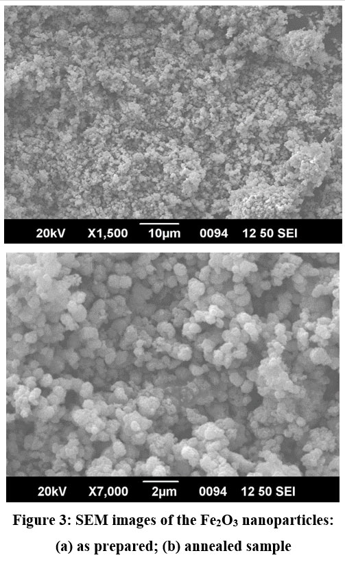 |
Figure 3: SEM images of the Fe2O3 nanoparticles: (a) as prepared; (b) annealed sample Click here to View Figure |
Antimicrobial Activity
The Fe2O3 nanoparticles were reported to have moderate antibacterial activity with MIC values of 100 ug/mL towards E. coli, S. aureus and S. pyogenus as well as 200 μg/mL towards P. aeruginosa as it was found in Table 1. Chloramphenicol, the classical antibiotic, presented lower values of MIC (50 µg/mL) against all the organisms tested, indicating that it is more potent. However, the results confirm that the ready to use Fe2O3 nanoparticles have enormous potential for use as antibacterial sources, and in particular when it comes to dealing with E. coli and S. pyogenus.
Table 1: Minimum inhibitory concentration of synthesized Fe2O3 nanoparticles against some bacterial strains
| Entry | E.coli | P. aeruginosa | S. aureus | S .pyogenus |
| MTCC 443 | MTCC 1688 | MTCC 96 | MTCC 442 | |
| Fe2O3 nanoparticles | 100 µg/mL | 200 µg/mL | 100 µg/mL | 100 µg/mL |
| Chloramphenicol | 50 µg/mL | 50 µg/mL | 50 µg/mL | 50 µg/mL |
| Antibacterial efficiency (%) | 50 | 25 | 50 | 50 |
The broth microdilution method was used to measure the antifungal activity of the prepared Fe2O3 nanoparticles, and this method was reported in our previous papers32. Nanoparticle suspensions were prepared with the help of DMSO being the solvent. The standard fungal strains (Candida albicans (MTCC 227), Aspergillus niger (MTCC 282) and Aspergillus clavatus (MTCC 1323)) were compared against standard fungi to determine the minimum inhibitory concentration (MIC) values.
Table 2 demonstrates that the Fe2O3 nanoparticles had a moderate antifungal activity with MIC values of 250 -1 g/mL against C. albicans and 500 -1 g/mL against A. niger and A. clavatus. Comparatively, the standard antifungal agents nystatin and griseofulvin had lower MIC values (100 µg/mL), which is more active. However, the results observed indicate that the Fe2O3 nanoparticles have a strong the potential of antifungal, which can be explained by their nanoparticle size, surface reactivity, and potential interaction with fungal cell membranes to cause growth inhibition.
Table 2: Minimum inhibitory concentration of synthesized Fe2O3 nanoparticles against some fungal strains
| Entry | C. albicans | A. niger | A. clavatus |
| MTCC 227 | MTCC 282 | MTCC 1323 | |
| Fe2O3 nanoparticles | 250 µg/mL | 500 µg/mL | 500 µg/mL |
| Nystatin | 100 µg/mL | 100 µg/mL | 100 µg/mL |
| Greseofulvin | 500 µg/mL | 100 µg/mL | 100 µg/mL |
| Antifungal efficiency(%) | 40 | 20 | 20 |
Antimalarial activity
Antimalarial activity of the synthesized Fe2O3 nanoparticles against Plasmodium falciparum cultures was tested and the values of antimalarial IC50 were obtained. As indicated in Table 3 the Fe2O3 nanoparticles show good antimalarial activity with an IC 50 of 0.80 μg/mL as compared to the standard drug quinine, which had an IC 50 of 0.268 μg/mL. The results showed a promising idea of the use of the nanoparticles as an antimalarial agent, despite the fact that Fe2O3 nanoparticles had less activity compared to the standard. This observed activity can be explained by the contact of nanoparticles with the parasitic cell membrane, which contributes to oxidative stress and damage to cellular metabolism. Increased surface area and the production of reactive oxygen species by Fe₂O₃ nanoparticles may be crucial for preventing the development of Plasmodium species. Their effectiveness and the potential for therapeutic use in treating malaria can be improved through further research to elucidate and optimize their structures.
Table 3: IC50 value of synthesized Fe2O3 nanoparticles against Plasmodium falciparum
| Entry | Name of compounds | Antimalarial activity
(IC50 μg/ml) |
Relative efficiency (%) |
| Fe2O3 nanoparticles | Fe2O3 nanoparticles | 0.80 μg/mL | 33.5 |
| Standard | Quinine | 0.268 μg/mL | 100 |
Conclusions
Co-precipitation of Fe2O3 was also achieved using a simple and affordable product co-precipitation technique that produced pure hematite phase with crystallites of nanoscale and spherical morphology. These nanoparticles have shown significant antimicrobial and antifungal properties against different pathogenic strains and this suggests that the nanoparticles have great potential in biomedical and environmental uses. The co-precipitation method was effective in the synthesis of Fe2O3 nanoparticles of high quality in mild conditions. Future studies must concentrate on the modification of surfaces in order to increase stability and biocompatibility and also to include expansion in the area of application, like catalysis, energy storage, and removal of contaminants in the environment.
Acknowledgment
The authors acknowledge the central instrumentation facility, SPPU, Pune, and the Sophisticated Test & Instrumentation Centre, Cochin University of Science and Technology, Cochin. The authors are grateful to the Microcare Research Laboratory, Surat, for antimicrobial evaluation.
Funding Sources
The author(s) received no financial support for the research, authorship, and/or publication of this article
Conflict of Interest
The authors do not have any conflict of interest
Data Availability Statement
This statement does not apply to this article
Ethics Statement
This research did not involve human participants, animal subjects, or any material that requires ethical approval
Permission to reproduce material from other Sources
Not applicable
Author Contributions
Naba Shaikh – conceptualization, methodology, data collection, Writing – Original Draft.
Nutan Sadgir – conceptualization, methodology, analysis, Writing Review & editing,
Sunil Dhonnar– writing manuscript, writing, Visualization, Supervision.
References
- Khan I, Saeed K, Khan I. Nanoparticles: Properties, applications and toxicities. Arabian Journal of Chemistry.Elsevier B.V. 2019;12(7):908-931. doi:10.1016/j.arabjc. 2017. 05. 011.
CrossRef - molecules-25-00112-v2.
- Elzein B. Nano Revolution: “Tiny tech, big impact: How nanotechnology is driving SDGs progress”. Heliyon.Elsevier Ltd. 2024;10(10). doi:10.1016/j.heliyon.2024.e31393
CrossRef - Jeevanandam J, Barhoum A, Chan YS, Dufresne A, Danquah MK. Review on nanoparticles and nanostructured materials: History, sources, toxicity and regulations. Beilstein Journal of Nanotechnology.Beilstein-Institut Zur Forderung der Chemischen Wissenschaften. 2018;9(1):1050-1074. doi:10.3762/bjnano.9.98
CrossRef - Cao L, Meziani MJ. Functional Nanocomposites: From Strategic Design to Applications. Nanomaterials.Multidisciplinary Digital Publishing Institute (MDPI). 2024;14(23). doi:10.3390/nano14231931
CrossRef - Ajaz M, Rasool W, Mahmood A. Comprehensive Review of Nanotechnology: Innovations and Multidisciplinary Applications. Futuristic Biotechnology. Published online March 31, 2024:12-18. doi:10.54393/fbt.v4i01.81
CrossRef - Bayda S, Adeel M, Tuccinardi T, Cordani M, Rizzolio F. The history of nanoscience and nanotechnology: From chemical-physical applications to nanomedicine. Molecules.MDPI AG. 2020;25(1). doi:10.3390/molecules25010112
CrossRef - Harish V, Tewari D, Gaur M, et al. Review on Nanoparticles and Nanostructured Materials: Bioimaging, Biosensing, Drug Delivery, Tissue Engineering, Antimicrobial, and Agro-Food Applications. Nanomaterials.MDPI. 2022;12(3). doi:10.3390/nano12030457
CrossRef - Akbarzadeh A, Samiei M, Davaran S. Magnetic nanoparticles: Preparation, physical properties, and applications in biomedicine. Nanoscale Res Lett. 2012;7. doi:10.1186/1556-276X-7-144
CrossRef - Salata O V. Applications of Nanoparticles in Biology and Medicine.; 2004. http://www.jnanobiotechnology.com/ content/2/1/3
CrossRef - Wang L, Liu H, Zhuang J, Wang D. Small-Scale Big Science: From Nano- to Atomically Dispersed Catalytic Materials. Small Science.John Wiley and Sons Inc. 2022;2(11). doi:10.1002/smsc.202200036
CrossRef - Nasrollahzadeh M, Atarod M, Jaleh B, Gandomirouzbahani M. In situ green synthesis of Ag nanoparticles on graphene oxide/TiO2 nanocomposite and their catalytic activity for the reduction of 4-nitrophenol, congo red and methylene blue. Ceram Int. 2016;42(7):8587-8596. doi:10.1016/j.ceramint.2016.02.088
CrossRef - Ajinkya N, Yu X, Kaithal P, Luo H, Somani P, Ramakrishna S. Magnetic iron oxide nanoparticle (Ionp) synthesis to applications: Present and future. Materials.MDPI AG. 2020;13(20):1-35. doi:10.3390/ma13204644
CrossRef - Cornell RMUS. The Iron Oxides: Structure, Properties, Reactions, Occurences and Uses. Vol John Wiley & Sons.; 2003.
CrossRef - Sivula K, Le Formal F, Grätzel M. Solar water splitting: Progress using hematite (α-Fe 2O3) photoelectrodes. ChemSusChem.Wiley-VCH Verlag. 2011;4(4):432-449. doi:10.1002/cssc.201000416
CrossRef - Wheeler DA, Wang G, Ling Y, Li Y, Zhang JZ. Nanostructured hematite: Synthesis, characterization, charge carrier dynamics, and photoelectrochemical properties. Energy Environ Sci. 2012;5(5):6682-6702. doi:10.1039/c2ee00001f
CrossRef - Auffan M, Rose J, Bottero JY, Lowry G V., Jolivet JP, Wiesner MR. Towards a definition of inorganic nanoparticles from an environmental, health and safety perspective. Nat Nanotechnol.Nature Publishing Group. 2009;4(10):634-641. doi:10.1038/nnano.2009.242
CrossRef - Tasnim NT, Ferdous N, Rumon MMH, Shakil MS. The Promise of Metal-Doped Iron Oxide Nanoparticles as Antimicrobial Agent. ACS Omega.American Chemical Society. 2024;9(1):16-32. doi:10.1021/acsomega.3c06323
CrossRef - Ashrafi-Saiedlou S, Rasouli-Sadaghiani MH, Fattahi M, Ghosta Y. Biosynthesis and characterization of iron oxide nanoparticles fabricated using cell-free supernatant of Pseudomonas fluorescens for antibacterial, antifungal, antioxidant, and photocatalytic applications. Sci Rep. 2025;15(1). doi:10.1038/s41598-024-84974-0
CrossRef - Gupta AK, Gupta M. Synthesis and surface engineering of iron oxide nanoparticles for biomedical applications. Biomaterials. 2005;26(18):3995-4021. doi:10.1016/j.biomaterials.2004.10.012
CrossRef - Wu W, He Q, Jiang C. Magnetic iron oxide nanoparticles: Synthesis and surface functionalization strategies. Nanoscale Res Lett. 2008;3(11):397-415. doi:10.1007/s11671-008-9174-9
CrossRef - Huber DL. Synthesis, properties, and applications of iron nanoparticles. Small. 2005;1(5):482-501. doi:10.1002/smll.200500006
CrossRef - Besenhard MO, Lagrow AP, Hodzic A, et al. Co-Precipitation Synthesis of Stable Iron Oxide Nanoparticles with NaOH: New Insights and Continuous Production via Flow Chemistry.
- Inestrosa-Izurieta MJ, Vilches D, Urzúa JI. Tailored synthesis of iron oxide nanoparticles for specific applications using a statistical experimental design. Heliyon. 2023;9(11). doi:10.1016/j.heliyon.2023.e21124
CrossRef - Farahmandjou M, Soflaee F. Synthesis and characterization of α-Fe2O3 nanoparticles by simple co-precipitation method. Physical Chemistry Research. 2015;3(3):191-196. doi:10.22036/pcr.2015.9193
- Lassoued A, Dkhil B, Gadri A, Ammar S. Control of the shape and size of iron oxide (α-Fe2O3) nanoparticles synthesized through the chemical precipitation method. Results Phys. 2017;7:3007-3015. doi:10.1016/j.rinp.2017.07.066
CrossRef - Hui BH, Salimi MN. Production of Iron Oxide Nanoparticles by Co-Precipitation method with Optimization Studies of Processing Temperature, pH and Stirring Rate. In: IOP Conference Series: Materials Science and Engineering. Vol 743. Institute of Physics Publishing; 2020. doi:10.1088/1757-899X/743/1/012036
CrossRef - Wiegand I, Hilpert K, Hancock REW. Agar and broth dilution methods to determine the minimal inhibitory concentration (MIC) of antimicrobial substances. Nat Protoc. 2008;3(2):163-175. doi:10.1038/nprot.2007.521
CrossRef - Kowalska-Krochmal B, Dudek-Wicher R. The minimum inhibitory concentration of antibiotics: Methods, interpretation, clinical relevance. Pathogens.MDPI AG. 2021;10(2):1-21. doi:10.3390/pathogens10020165
CrossRef - Kadeřábková N, Mahmood AJS, Mavridou DAI. Antibiotic susceptibility testing using minimum inhibitory concentration (MIC) assays. npj Antimicrobials and Resistance. 2024;2(1). doi:10.1038/s44259-024-00051-6
CrossRef - Sadgir N V., Dhonnar SL, Jagdale BS. Synthesis, molecular structure, FMO, spectroscopic, antimicrobial and in-silico investigation of (E)-1-(benzo[d][1,3]dioxol-5-yl)-3-(4-aryl)prop-2-en-1-one derivative: Experimental and computational study. Results Chem. 2023;5. doi:10.1016/j.rechem.2023.100887
CrossRef - Sadgir N V., Adole VA, Dhonnar SL, Jagdale BS. Synthesis and biological evaluation of coumarin appended thiazole hybrid heterocycles: Antibacterial and antifungal study. J Mol Struct. 2023;1293. doi:10.1016/j.molstruc.2023.136229
CrossRef
Accepted on: 30 Jan 2026
Second Review by: Dr. Suresh Kumar Lakhanpal
Final Approval by: Dr. Shouxun Ji


![]()
![]()
![]()
![]()
