Phase formation and Morphological features of Calcium Copper Titanate by Modified Solid State Process
Ashnarayan Sah1, Soumya Mukherjee2* , Mohammed Shahnawaz3, Sath Banerjee1
1Department of Metallurgical and Materials Engineering, Jadavpur University, Kolkata-700032, India
2Department of Metallurgical and Materials Engineering, Kazi Nazrul University, Asansol-713340, India
3Department of Electronics and Telecommunication Engineering, Heritage Institute of Technology, Kolkata-700107, India
Corresponding Author E-mail: smmukherjee3@gmail.com, smmukherjee4a@gmail.com
DOI : http://dx.doi.org/10.13005/msri/160105
Article Publishing History
Article Received on : 07-03-2019
Article Accepted on : 12-04-2019
Article Published : 12 Apr 2019
Plagiarism Check: Yes
Reviewed by: Jagadeesh S
Second Review by: Gauri Khanolkar
Final Approval by: Jit Satyabrata
Article Metrics
ABSTRACT:
Perovskite calcium copper titanate posses giant dielectric constant making it a suitable candidate for possible applications in microelectronic components, advanced transistors, energy storage capacitors. Generally, this grade of material is synthesized by the chemical route to improving homogeneity, controlled size growth for enhanced properties. In the present research, a simple synthesis process was adopted using precursors of high purity oxides like Calcium carbonate, titania, Copper oxide without any use of complicated synthesis routes and costly chemical precursors. The molar ratio of oxides used was about 1:3:4 with mechano-chemical activation in an agate mortar for 20, 25 and 30 hours respectively in dry condition. After milling, powders obtained were made to undergo annealing at a fixed temperature of 900°C for 26 hours soaking period. Phase analysis was carried to determine the phase along with crystallite size calculation. Bonding information of the synthesized sample was analyzed to obtain the M-O coordination and vibration-stretching analysis of the bonds. Morphological features were also noted using FESEM (Field Emission Scanning Electron Microscopy) for understanding grains and granular boundaries. Both FTIR (Fourier Transform Infra-Red Spectroscopy) and XRD (X-Ray Diffractogram) analyses confirm the compound formation in terms of molecular structure responsible to obtain the proper phase.
KEYWORDS:
Calcium Copper Titanate; FTIR; Morphology; XRD
Copy the following to cite this article:
Sah A, Mukherjee S, Shahnawaz M, Banerjee S. Phase formation and Morphological features of Calcium Copper Titanate by Modified Solid State Process. Mat. Sci. Res. India; 16 (1).
|
Copy the following to cite this URL:
Sah A, Mukherjee S, Shahnawaz M, Banerjee S. Phase formation and Morphological features of Calcium Copper Titanate by Modified Solid State Process. Mat. Sci. Res. India; 16 (1). Available from: https://bit.ly/2VGmFmv
|
Introduction
Perovskite having ABO3 formula is a versatile ceramic structure with applications as semiconductor, catalyst, sensors, memory devices and others. Calcium Copper titanate is a modified perovskite structure material (also referred to as aurivillius structure) having applications in the above-mentioned domain in addition to application as magnetic materials after doping. The parent structure of perovskite is modified due to superimposition of body-centred ordering of Ca, Cu along with prominent tilting of Titanium centred octahedral.1 The ceramic is also known as colossal relative permeability due to the high dielectric constant of about 10.4 High dielectric constant also influences the volume of the electrical devices in applications. The origin for large dielectric constant is still under investigations. It has been reported in research studies that TiO2 and perovskite oxide lose oxygen during high-temperature sintering while reoxidation occurs during cooling. During cooling, re-oxidation occurs at the surface layers and at the grain boundaries as a cause of rapidly falling temperature and availability of short time. As a consequence, insulating layers are formed at surface and grain boundaries while the bulk grain remains semiconducting in nature.2 The giant dielectric constant of the material is found to be almost independent of temperature in the range 100-400K. Chung et al. observed a non-linear dielectric coefficient in the ceramic perovskite to be about 900 quite higher than ZnO which is used as a classical varistor. Calcium copper titanate also exhibits high dielectric loss limiting its application in the domain of devices. This hindrance of the material is made to overcome by undergoing substitution at the proper site. The scientific reason behind such suppression of dielectric loss may be due to retardation of barrier layer formation at grain boundaries. The material is noted to follow non-ohmic characteristics at such conditions.3 The mentioned perovskite is observed to be synthesized by sol-gel process1,4,5,6 mechanochemical process7,8 solid-state processing9, 10 hydrothermal11 and others. Some other popular methods of synthesis are combustion synthesis method or self-propagating high-temperature synthesis, sonochemical assisted and co-precipitation technique. Calcium copper titanate is noted to be applied for capacitors, antennas, microwave devices and sensors. The porous polycrystalline nanostructure is found to be quite beneficial for sensor applications. One of the significant properties of Calcium Copper titanate-based sensor is the ability to detect, monitor gases and toxic species without decomposition or changes in their molecular arrangement.12 In the present article, the modified solid-state process is adopted to synthesize utilizing proper molar ratio. The synthesized material is analyzed for phase determination by XRD, stretching bonding and M-O co-ordinations by FTIR analysis while morphology is studied by FESEM.
Experimental Procedure
In the synthesis of perovskite (Aurivillius) Calcium Copper Titanate, Calcium Carbonate, Cupric Oxide and titania (AR Grade, Merck India) were selected as precursors. All the powders were taken in a proper stoichiometric ratio of CaCO3:CuO: TiO2 as 1:3:4 and made to undergo milling activation in arid condition. Milling activation was carried for 20 hours, 25 hours and 30 hours respectively followed by annealing at 900°C for 26 hours. Phase analysis was carried by XRD (Rigaku, Ultima III, Cu Kα1.54A°, 50KV, 40mA) for synthesized powder samples. Scan range for the sample was 10 to 90° with a scan rate of 5°/min along with the determination of crystallite size and planes of orientation. Morphology of the sample was studied using FESEM, Hitachi S-4800 in a powder state. The sample was placed on carbon tape with conducting coating over it to prevent static charge accumulation for better resolution and imaging. Bonding and metal coordination analysis was carried for powder samples by FTIR analysis (IR Prestige -21, Shimadzu) after mixing it with KBr for pellet preparation. Pellets were prepared for FTIR analysis by undergoing pressing under pressure of 6t/cm2 in a hydraulic press.
Results and Discussions
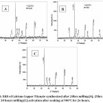
Figure 1: XRD of Calcium Copper Titanate synthesized after 20hrs milling (A), 25hrs milling (B) and 30 hours milling (C) activation after soaking at 900°C for 26 hours.
Above figures execute XRD of the synthesized samples. In all cases, sharp intense peaks are noted without any consultation or peak splitting. Intense sharp peaks are indications of crystalline nature of the synthesized materials. There is no trace of diffuse peaks or humps in the material matrix. The XRD patterns confirm the single phase structure. All three samples are made to undergo three different milling time variations but the same soaking period at a constant temperature of about 900°C is maintained. At this fixed composition, sharp peaks are noted within the 2θ scan range at 34.55°, 38.8°, 49.50° and 61.65° respectively. Small peaks can also be observed about 2θ values of 27.75°, 35.80°, 42.60° and 75.30° respectively. The peaks are indexed as per the (hkl) planes reported in JCPDS file (75-1149). The appearance of peaks corresponding to the CCTO phase indicates the formation of a single phase compound. The above XRD peaks are in correspondence with findings by N. Tripathy et al,13 R.E.-Gonza´lez et al.,7 where synthesis is carried by solid state routes followed by a sputtering and mechanochemical process. Some minor impurities like copper oxide are noted at about 35.88° after 20 hours milling, 26 hours annealing at 900ºC. Similarly, for 25 hours milling, minor peaks of impure oxides of Ti is noted respectively. For 30 hours milling, the sample is noted to be the purest without any formation of other metal oxides instead of desired perovskite compound. Crystallite size obtained by Scherrer’s formula is noted to be about 84.47nm, 82.66nm for 20 hrs, 30 hours sample and around 122 nm for 25 hours mill sample. The sudden change may be due to the formation of TiO as noted from XRD analysis. TiO is metastable in nature formed as intermediate during milling which later converts to perovskite after milling reaction followed by annealing. Due to the development of minor secondary phase, nucleation, the growth of phases undergo metal stabilization and hence such possible drastic increase in crystallite size happens. Moreover, 30 hours milling followed by 26 hours heat treatment leads to pure calcium copper titanate without any presence of other oxides as noted from XRD analysis with reference to JCPDS of Titanium oxide (81-1286), Copper Oxide (80-1917) used as precursors.
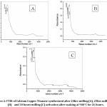
Figure 2: FTIR of Calcium Copper Titanate synthesized after 20hrs milling (A), 25hrs milling (B) and 30 hours milling (C) activation after soaking at 900°C for 26 hours.
Figure 2 represents the FTIR spectra of the synthesized samples. Major peaks of vibration for M-O co-ordinations are noted within 1000cm-1 indicating the successful synthesis of the inorganic structure. Absorption peaks are noted at about 530cm-1, 450cm-1, 500cm-1 mainly. The above absorptions are corresponding to Ca-O, the vibration mode of the O-Ti and bending of Cu-O bonds present in the synthesized samples. The experimental characteristics bands are noted within the scan range of 380cm-1 to 700cm-1 encompassing the mixture of CuO4 and TiO6 groups. The above experimental bands and peaks indicate the presence of CCTO as sample validating the XRD phase determination. The above experimental findings are in correspondence with the results of P. Thirumanathan et al who have studied optical, electrical properties of sol-gel synthesized calcium copper titanate powders.5 Almeida observed three main absorptions at 561, 516, 437cm-1 and Ti-O vibrations at about 653-550cm-1 while for Ti-O-Ti it is about 495-436cm-1.14 Thus above findings are noted to be in correspondence with research article by Almeida et.al which focussed on mechanochemical alloying of Calcium copper titanate.
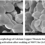
Figure 3: FESEM morphology of Calcium Copper Titanate formed after 30 hours of milling activation after soaking at 900°C for 26 hours.
FESEM morphology of the best synthesized conditioned sample is noted after confirming the s from phase analysis and FTIR analysis on bonding, M-O information. From XRD analysis it is concluded that 30 hours milling followed by 26 hours soaking period at 900ºC yields the purest perovskite. Morphology of the synthesized sample at the above-mentioned conditions exhibits proper granular formation with strong agglomeration tendency. Few minute pores are noted within the agglomerated form. Strong interconnections among grains are noted. Grains are having a polygonal shape with conchoidal fracture face at the top while some are having a spherical morphology. Individual grains are around 500nm to about 200nm, 100nm in some cases. Grains are found to get interlocked in a particular fashion leading to floral one with conchoidal fracture granules around the central hub of the agglomerates. There is an absence of any unwanted distortion observed in the matrix of the synthesized sample.
Conclusions
Modified Solid State Process was utilized to prepare modified perovskite structure material Calcium Copper titanate. Crystallite size was noted to be about 84.47nm, 82.66nm for 20 hrs, 30 hours sample and around 122 nm for 25 hours mill sample. Bonding and stretching vibration analysis of M-O coordination were noted from FTIR analysis. FESEM analysis reveals the morphology to be granular in nature with strong agglomeration, minute porosity while grains were a polygonal shape with conchoidal fracture.
Acknowledgement
Corresponding would like to thank HOD, Department of Metallurgical & Materials Engineering for providing XRD facilities to study phase formation. The author also acknowledges Prof (Dr.) Kalyan Kumar Chappopadhyay, Director, SMSNT, Jadavpur University for providing FTIR and FESEM facilities.
Funding Sources
There are no funding sources for carrying the experiment in the laboratory.
References
- Jesurani S., Kanagesan S., Velmurugan R., Kalaivani T., Phase formation and high dielectric constant of calcium copper titanate using the sol-gel route. J. Mater. Sc. Electron. (2012); 23: 668-674.
CrossRef
- Rai A. K., Singh N. K., Lee S-K., Mandal K.D., Kumar D., Om Parkash, Dielectric properties of iron-doped calcium copper titanate, CaCu2.9Fe0.1Ti4O12. J. Alloy. Compd. (2011); 509: 8901-8906.
CrossRef
- Chung S. Y., Kim Il. D., Kang S.J.L., Strong nonlinear current-voltage behaviour in perovskite derivative calcium copper titanate. Nat. Mater. (2004); 3: 774-778.
CrossRef
- Xu D., He K., Chen B., Xu Ch., Sh. Mua, Jiao Lei., Sun Xi., Yang Y., Microstructure and electric characteristics of AETiO3 (AE=Mg, Ca, Sr) doped CaCu3Ti4O12 thin films prepared by the sol-gel method. PRO. NAT. SCI-MATER. (2015); 25: 399-404.
- Thiruramanathan P., Marikani A., Madhavan D., Optical and Electrical Properties of Sol-Gel Synthesized Calcium Copper Titanate Nanopowders, Int. J. Chem. Tech. Res. (2015); 8, No. 3, 981-987.
- Jesurani S., Kanagesan S., Velmurugan R., Thirupathi C., Sivakumar M., Kalaivani T., Nanoparticles of the giant dielectric material, Calcium Copper titanate from a Sol-gel technique. Mater. Lett. (2011); 65: 3305-3308.
CrossRef
- -Gonzalez R.E., Vega E., Tamayo R., Criado J.M., Dia´nez M. J., Mechanical Processing of CaCu3Ti4O12 with Giant Dielectric Properties. Mater. Manuf. Processes. (2014); 29: 1179-1183.
CrossRef
- Alizadeha M., Ardakani H.A., Amini R., Ghazanfari M.R., Ghaffari M., Structural and phase evolution in mechanically alloyed calcium copper titanate dielectrics. Ceram. Int. (2013); 39: 3307-3312.
CrossRef
- Ramirez A.P., Subramanian M.A., Gardela M., Blumberga G., Li D., Vogt T., et al., Giant dielectric constant response in a copper-titanate. Solid. State. Commun. (2000); 115: 217–220.
CrossRef
- Yu H., Liu H., Hao H., Guo L., Jin C., Yu Z., et al., Grain size dependence of relaxor behaviour in CaCu3Ti4O12 ceramics. Appl. Phys. Lett. (2007); 91: pp 222911 (22).
- Oliveira L.H., de Moura A.P., Mazzo T.M., Ramírez M.A., Cavalcante L.S., Antonio S.G., Avansi W., Mastelaro V.R., Longo E., Varela J.A., Structural refinement and photoluminescence properties of irregular cube-like (Ca1-xCux) TiO3 microcrystals synthesized by the microwave hydrothermal method. Mater. Chem. Phys. (2012); 136: 130-139.
CrossRef
- Ahmadipour M., Ain M.F., Ahmad Z. A., A Short Review on Copper Calcium Titanate (CCTO) Electroceramic: Synthesis, Dielectric Properties, Film Deposition and Sensing Application. Nano-Micro Lett. (2016); 8: 291-311.
CrossRef
- Tripathy N., Das K. C., Ghosh S. P., Bose G., Kar J. P., Fabrication of high-k dielectric Calcium Copper Titanate (CCTO) target by solid state route. IOP Conf. Series: Materials Science and Engineering (2016); 115: 012022 doi:10.1088/1757-899X/115/1/012022
CrossRef
- Almeida A.F.L., De Oliveira R.S., Go´es J.C., Sasaki J.M., Souza Filho A.G., Mendes Filho J., Sombra A.S.B., Structural properties of CaCu3Ti4O12 obtained by mechanical alloying. Mater. Sci. and Engg. B. (2002); 96: 275-283.
CrossRef

This work is licensed under a Creative Commons Attribution 4.0 International License.