Effect of Mn Dopant on Structural and Optical Properties of NiFe2O4 Nanoparticles Synthesized by Autocombustion Method.
M. B. Shelar1*, K. M. Patil 2, S. K. Nalugade2, V. R. Chavan2, R. M. Jadhav2, M. S. Kore2 and A. V. Gaikwad2
1Materials Research Laboratory, Department of Physics , D. Y. Patil College of Engineering and Technology (An Autonomous Institute), Kasaba Bawada, Kolhapur-416006, Maharashtra, India
2Department of Chemical Engineering, D. Y. Patil College of Engineering and Technology (An Autonomous Institute), Kasaba Bawada, Kolhapur-416006, Maharashtra, India
Article Publishing History Article Received on : 12 Mar 2024 Article Accepted on : 17 Apr 2024 Article Published : 23 Apr 2024 Plagiarism Check: Yes Reviewed by: Dr. M. Narasimha Murthy Second Review by: Dr. Oluwatoyin Olasanmi Final Approval by: Dr. K. M. Garadkar
Article Metrics
ABSTRACT:
The work is done on the Structural and optical properties of Manganese doped nickel nano ferrites prepared by auto route. The manganese is doped in nickel (Mn-Ni) mixed nano ferrites MnxNi1-xFe2O4 (x = 0.0, 0.2, 0.4, 0.6, 0.8, 1.0) have been synthesized by a simple auto combustion route. The cubic spinel structure were confined by using X- ray Diffraction for MnxNi1-xFe2O4 nano ferrite particles which shows favored positioning along with (311) direction. However, size of crystal increases from 20 nm to 26.38 nm increase in Mn content. The optical properties were examined by means of UV-visible absorption spectroscopy observed from 200 to 800 nm. Using the absorption spectra and Tauc’s relation, the band gap was calculated and varies between 3.57 and 3.80 eV. The significant decline in the optical band gap was calculated with increasing manganese Composition from 3.80ev to 3.57 eV up to Mn=0.6, The further increase due to preparative crystallinity defects in composition from 3.67eV to 3.70 eV shows tunability of the optical band gap varying the concentration of Mn in nano ferrites. The present nanoparticles with variable narrow band gap may find applications in photocatalysis to increase the efficiency of photocatalytic reactions.
KEYWORDS:
Auto combustion route; Mn-Ni ferrite; Structural properties; Optical properties
Copy the following to cite this article:
Shelar M. B, Patil K. M, Nalugade S. K, Chavan V. R, Jadhav R. M, Kore M. S, Gaikwad A. V. Effect of Mn Dopant on Structural and Optical Properties of NiFe2O4 Nanoparticles Synthesized by Autocombustion Method. Mat. Sci. Res. India;21(1).
Copy the following to cite this URL:
Shelar M. B, Patil K. M, Nalugade S. K, Chavan V. R, Jadhav R. M, Kore M. S, Gaikwad A. V. Effect of Mn Dopant on Structural and Optical Properties of NiFe2O4 Nanoparticles Synthesized by Autocombustion Method. Mat. Sci. Res. India;21(1). Available from: https://bit.ly/4dcmuaI
Introduction
Nano ferrite particles are recently finding potential applicant in automated industries due to their desirable optical and electrical properties1-2. Due to different properties of ferrites it has been prepared to meet the remarkable uses in microwave fields, engineering, permanent magnet, transformer core and memory chips etc. The spinel ferrites are semiconducting in nature. The electrical property depends on several factors including the method of preparation, sintering temperature, sintering atmosphere, chemical composition and microstructure. as well as, by addition of impurities3. Ferrites containing Mn2+ions tend to form α-Fe2O3 phase when heat treated above 200 ℃ in air atmosphere4. It is very important to understand the mechanism involve in the change of properties. Nano ferrites are useful in types of biomedical fields such as X-ray diagnosis, drug delivery, hyperthermia, and magnetic resonance imaging (MRI)5-8. Manganese ferrite (MnFe2O4) nanoparticles (NPs) with other ferrites are considered as a crucial tool for enhancing efficiency of magnetic resonance imaging, hyperthermia, and drug delivery9. Also, it is a mixed ferrite, where Mn2+ and Fe3+ occupies both tetrahedral and octahedral bonding sites10. The chemical changes in ferrites depend on their structural parameters of particle size and shape, which can be modified in the synthesis processes. In spinel ferrites, the physical and magnetic properties are powerfully reliant on cation distribution and process of research 11,12.
In the present work, the studies on MnxNi1−xFe2O4 (x = 0,0.2,0.4,0.6,0.8,1.0) ferrites annealed at 600 ℃ are reported. Powder X-ray diffraction (XRD), FT-IR spectroscopy techniques and spectrophotometer stayed active for structural and optical characterizations.
Material and methods
Materials
For preparation of Mn doped Nickel nanoparticles, stoichiometric amounts of AR grade Manganese (II) Nitrates (Mn (NO₃)₂ .6H2O, (purity ~ 99%), AR grade Iron (III) Nitrates (Fe(NO₃)₃. 9H2O, (purity~ 98%) and AR grade Nickel (II) Nitrates (Ni(NO₃). 6H2O), (purity ~ 99%) were used as starting materials and PVA (Polyvinyl Alcohol) and sucrose solutions from Merck, Mumbai were used as precipitating agent. Deionized water was used as solvent.
Sample Preparation
Manganese doped Nickel ferrite nanoparticles were prepared by self-propagating auto combustion route from a mixture of stoichiometric amount of Mn (NO₃)₂ .6H2O, Fe(NO₃)₃. 9H2O and Nickel Nitrates (Ni(NO₃).6H2O). 10% of PVA and sucrose solutions were added it, where the sucrose is utilized for the combustion purpose and PVA forms the polymer resin with metal ions trapped in it. The whole mixture was heated to about 80oC for the evolution of NO2, CO2, and H2O. The mixture in solution transforms and converted to black fluffy gel. The PVA (the matrix) and Sucrose (the fuel) are dissolved together and at elevated temperature, and the brown fumed of solution gets evaporated. Sucrose provides the wrapping throughout the coordination for the cations in solution and circumvents their selective precipitation during the evaporation process. Black fluffy, and voluminous gel gets burnt in the self-propagating manner at about 90 ℃. for 6 h to remove In order of residual water, the as-prepared Nickel ferrite nanoparticles was dried overnight and calcined at 600 ℃. The prepared samples were used to study the structural and optical characterisation.
Characterization
Structural properties of the prepared MnxNi1-xFe2O4 (x = 0, 0.2, 0.4, 0.6, 0.8 and 1.0) nano ferrites were observed using the X-ray diffractometer (Rigaku Miniflex 600 with Cu Kα1 radiation at 40 kV, 25 mA over the 2θ range of 10–100o (λ = 1.54056 Å). The optical properties of Nano powders were studied using the UV–Vis. Spectrophotometer (JASCO- V-770).
Results
Result and Discussion
Structural analysis
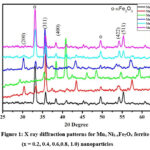
Figure 1: X ray diffraction patterns for Mnx Ni1-xFe2O4 ferrite (x = 0.2, 0.4, 0.6,0.8, 1.0) nanoparticles
The XRD pattern of the prepared samples is shown in Fig 1 with noticeable (hkl) planes of (200), (311), (400), (422), (511), and (440) which are indexed with JCPDS card nos. 00-019-0629 for Fe3O4 and 00-010-0319 for MnFe2O4. The well-defined diffraction patterns confirm that the chemical reaction is completed. The observed diffraction peaks correspond to cubic spinel phase with the addition of some minor peaks as secondary phase (α-Fe2O3). The intensity of these peaks gently increases with an increase in the concentration of Mn2+ ion.
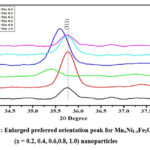
An enlarged view of the high-intensity characteristic peak (311) as shown in Fig.2. Manganese ions may have different ionic radius compared to nickel ions, leading to change in the lattice parameter of crystal structure, effect on spacing between lattice planes influencing the shift in position of (311) peak. It can induce strain in the crystal lattice, altering the diffraction pattern and casing peak shifts 13.
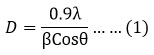
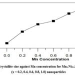
The average crystallite size of the resulting powder materials, calculated using the Debye-Scherrer formula (equation 1), increased monotonically with increasing Mn concentration (0.0-1.0) from 20.0 to 26.38 nm, respectively 14 as shown in Fig.3.
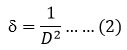
X-ray wavelength, FWHM in radians and Bragg’s angle, respectively, refer to λ, β, and θ. and dislocation density (d) as
Also, the larger ionic radius of Mn2+ compared to Ni2+ leads to stronger ionic interactions between them15. An increase in crystallite size typically results sharper and narrower peaks in an XRD pattern. This is because larger crystallites lead to a decrease in width of diffraction peaks, which enhances resolution of XRD pattern. So, for Mn doped nickel ferrites, sharp and crystalline peaks are observed after doping. The dislocation density decreases with increase in Mn concentration reveals that the less distortion in crystal lattice.
Figure 3: Crystallite size against Mn concentration for Mnx Ni1-xFe2O4 ferrite (x = 0.2, 0.4, 0.6, 0.8, 1.0) nanoparticles
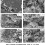
The SEM micrographs for ferrite phase i.e. Mnx Ni1-xFe2O4 ferrite (x = 0.2, 0.4, 0.6,0.8, 1.0) composition is shown in Fig.4. The grains in the ferrite phase are smaller in size. The uniform nature of the particles is revealed with some agglomeration. The average grain size is about 97 nm. There is progress of grain size with calcination temperature. Increasing the Mn2+ ion concentration, the powder show irregular microstructures with small spherical particles and size of the particle is varied [16]. Generally, the grain size increases with an increase in Mn2+ ion concentration.
Figure 4: Scanning Electron Micrographs for MnxNi1-xFe2O4 ferrite (x = 0.2, 0.4, 0.6,0.8, 1.0) composition
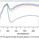
UV-Visible Spectroscopy is used for the examination of the optical properties of Mn doped Ni ferrite nanoparticles. Solutions of Mn-Ni nano ferrites prepared by sonication in double distilled water were used to record the spectra. The absorption spectra were observed from 200nm to 800 nm as shown in figure Fig.5.
From absorption spectra, the variation in Mn composition in Ni ferrites causes the absorption change in the UV section about 300 nm (for Mn0.8Ni0.2Fe2O4) moves to longer wavelength visible region. This shifting to longer wavelength can be attributed to changes in electronic structure and composition of the material. Manganese doping alters the energy level and band structure, affecting absorption characteristics and again increase in absorption wavelength. To examine influence of Manganese effect on optical band gap, the Tauc equation is used for fitting absorption data17.
where C is proportionality constant, h is Planck constant, Eg is band gap, n is an integer which is ‘2’ for direct band gap transition in present study and α is the absorption coefficient.
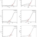
The plot of (ahν)2 with respect to energy of photon (hν) for all the Mn doped nickel nano ferrites are shown in Fig. 6. The extrapolation of the curve to the energy axis gives the band energy value for particular composition.
Figure 6:Plot of (ahν)2 against hν for the prepared Mn0.0Ni1.0Fe2O4 (x= 0.0, 0.2, 0.4, 0.6, 0.8, 1.0) nanoparticles.
The values of band gap vary from 3.80 eV – 3.57 eV for Mn doped nickel ferrites. Thus, up to Mn=0.6 the projected value of band gap decreases is due to the quantum confinement effect. Further for Mn = 0.8 and 1.0 composition, the band gap rises may due to the little disorder in crystallinity of compositions. The increase in Mn produces additional energy levels in band structure. These levels can hybridize with existing energy bands of material, altering the band after Mn =0.8. Thus addition of Manganese for nickel ferrite leads to changes in electronic properties of materials in terms of tuned band gap, conductivity and absorption spectra.
Conclusions
Manganese doped nickel nanoparticles was synthesized by using self-propagating auto combustion method. The cubic spinel structure of Mn-Ni nanoparticles was identified by XRD. The agglomerated nature of ferrite particles nature is confirmed by Scanning electron Microscopy. The result indicate that the structural properties of nickel ferrite can be improved by Mn addition. The optical band gap for Mn- Ni nanoparticles decreases from 3.80 to 3.57 eV up to Mn = 0.6. The further rise of band gap only because of preparative crystallinity defects in composition revealed the variation of Mn in Ni ferrites for tuning its band gap. Thus variable narrow band gap can increase the efficiency of photo catalysis by enabling the absorption of a wide range of photons from solar spectrum and enhancing the generation of electron-hole pairs and facilitating more photocatalytic reactions.
Acknowledgement
The authors wish to thank Department of Physics, Shivaji University, Kolhapur for XRD measurements, D. Y. Patil Medical College, Kolhapur for FTIR measurement and Jaysingpur College, Jaysingpur (Department of Physics), Kolhapur for UV-visible spectroscopy measurement
Conflict of Interest
None
Funding Source
None
References
Manikandan A, Vijaya JJ, Kennedy LJ, Bououdina M. “Structural optical and magnetic properties of Zn1-xCuxFe2O4 nanoparticles prepared by microwave combustion method”, J. Molecular Str. 2013; 40: 1035:332 CrossRef
Gopalan E.V., Malini K.A., Sagar S, Kumar D.S., Yoshida Y, Al-Omari I.A, Anantharaman M.R. “Mechanism of ac conduction in nanostructured manganese zinc mixed ferrite”, J. Phys. D: Appl. Phys., 2009(16) :165005 CrossRef
Devi E.C., Soibam I. “Effect of Zn doping on the structural, electrical and magnetic properties of MnFe2O4 nanoparticles”. Indian J. of Phys., 2017: 1-7 CrossRef
E. Ranjith kumar , R. Jayaprakash , M.S. Seehra , T. Prakash , Sanjay Kumar, “Effect of a-Fe 2 O3 phase on structural, magnetic and dielectric properties of Mn–Zn ferrite nanoparticles” J. Phys. Chem. Solid.:2013: 74(7):943–949 CrossRef
Valenzuela R., “Novel applications of ferrites,” Physics Research International, vol., Article ID 2012: 2012 (9) :591839 CrossRef
Latorre-Esteves M., Cortés A., Torres-Lugo M. and Rinaldi C., “Synthesis and characterization of carboxymethyl dextran-coated Mn/Zn ferrite for biomedical applications,” J. Magn. Mag. Mater, 2009: 321(19) 3061–3066 CrossRef
A. Nigam and S. J. Pawar, “Structural, magnetic, and antimicrobial properties of zinc doped magnesium ferrite for drug delivery applications,” Ceram. Inter. 2020: 46 (4) : 4058–4064 CrossRef
Kefeni K. K., Msagati T. A. M., Nkambule T. T. I. Mamba B. B., “Spinel ferrite nanoparticles and nanocomposites for biomedical applications and their toxicity,” Mater. Sci. Engin.: C, 2020: 107: 110314 CrossRef
Javed F., Abbas M. A, Asad M. I. et al., “Gd3+ doped CoFe2O4 nanoparticles for targeted drug delivery and magnetic resonance imaging,” Magnetochemistry, 2021: 7 (4): 47 CrossRef
Ju W., Park J. H., Jung H. R., Cho S. J. and Lee W. J., “Electrospun MnFe2O4 nanofibers: preparation and morphology,” Comp. Sci. Techn. 2008: 68 (7-8) : 1704–1709, CrossRef
Maaz, K., Karim, S., Mumtaz, A., Hasanain, S.K., Liu J., Duan J.L., “Synthesis and magnetic characterization of nickel ferrite nanoparticles prepared by co-precipitation route”, Magn. Magn. Mat. 2009: 321: 1838–1842 CrossRef
Hui, D.C., Xue, W.G., Lei, S., Wei, G.D., Jun, J.C., Sheng, X.D. “Investigation of the thermal stability of Mn ferrite particles synthesized by a modified co-precipitation method”, Sci. China Phys. Mech. Astron. 2013:56(3): 568–572 CrossRef
Md. M. Roni, K. Hoque, A. C. Paul , M. N. I. Khan Synthesis of La-doped Mn0·6Zn0.4LaxFe2-xO4 and the study of its structural, electrical and magnetic properties for high frequency applications, Results in Materials 11:100215 CrossRef
P. Monisha , P. Priyadharshini , S.S. Gomathi , K. Pushpanathan, “Influence of Mn dopant on the crystallite size, optical and magnetic behaviour of CoFe2O4 magnetic nanoparticles”, J. Phys. Chem. Solid., 2021: 148: 109654 CrossRef
Gosh PK, Ahmed SF, Jana S, Chattopadhyay K.K., “Photoluminescence and field emission properties of ZnS:Mn nanoparticles synthesized by rf-magnetron sputtering technique”, Optical Mater., 2007 : (12) :1584-90. CrossRef
Ranjith Kumar, E., Siva Prasada Reddy, P., Sarala Devi, G., Sathiyaraj, S.: Structural, dielectric and gas sensing behavior of Mn substituted spinel MFe2O4 (M = Zn, Cu, Ni, and Co) ferrite nanoparticles. Magn. Magn. Mat. 2016: 398, 281–288 CrossRef
Tahir, M.B., Iqbal, T., Hassan, A., Ghazal, S. “Wet chemical Coprecipitation synthesis of nickel ferrite nanoparticles and their characterization”, J. Inorg. Organomet. Polym. Mater. 2017: 27, 1430– 1438 CrossRef

 , V. R. Chavan2, R. M. Jadhav2