Synthesis, Characterization and Thermal Studies of a N, N’- bis(2- Hydroxy- Alpha- Methyl Benzylidene) -1, 9- Nonanyldiamine Uranyl (VI) nitrate [UO2(HMND)]2+
Shahriar Ghammamy1* and Sajjad Sedaghat2
1Department of Chemistry, Faculty of Science, Imam Khomeini International University, Qazvin, Iran.
2Department of Chemistry, Faculty of Science, Islamic Azad University, Malard Branch, Malard, Iran.
DOI : http://dx.doi.org/10.13005/msri/090201
Article Publishing History
Article Received on : 20 Feb 2012
Article Accepted on : 25 Mar 2012
Article Published :
Plagiarism Check: No
Article Metrics
ABSTRACT:
N, N’- bis(2- hydroxy- alpha- methyl benzylidene) -1,9- nonanyldiamineabbreviated as HMND was synthesized and characterized. N, N’- bis(2- hydroxy- alpha- methyl benzylidene) -1,9- nonanyldiamineUranyl (VI) nitrate prepared by reaction of nitrate salt of UO2(NO3)2.6H2O with HMND. In this research, some of the inorganic complexes of uranyl with N- donor ligands were synthesized. Complexes were characterized by FT-IR and UV, ¹HNMR, ¹³CNMR spectra, TG/DTG measurements and some physical properties. The results of simultaneous TG-DTG-DTA analyses of the complexes show the final degradation product for these complexes are UO3. The antitumor activity of used ligands and their complexes against a panel of human tumor cell lines (HT29: Haman colon adenocarcinoma cell line T47D: human breast adenocarcinoma cell line) were studied and determined by MTT (3-[4,5-dimethylthiazol-2-yl]-2,5-diphenyl-tetrazolium bromide) assay. These data suggest that some of these compounds provide good models for the further design of potent antitumor materials. Also the results show chelation causes drastic change in the biological properties of the ligands and also the metal moiety. So the toxic effects of uranyl can be prevented by using chelating agent and complexation of the potentially multidentate ligands.
KEYWORDS:
N; N’- disalicylidene -p- chlorobenzyldiamineUranyl (VI) nitrate; Synthesis; Thermal analysis; FT-IR and UV–visible spectroscopy; Schiff bases, Anticancer activity
Copy the following to cite this article:
Ghammamy S, Sedaghat S. Synthesis, characterization and Thermal Studies of a N, N’- bis(2- hydroxy- alpha- methyl benzylidene) -1, 9- nonanyldiamine Uranyl (VI) nitrate [UO2(HMND)]2+. Mat.Sci.Res.India;9(2)
|
Copy the following to cite this URL:
Ghammamy S, Sedaghat S. Synthesis, characterization and Thermal Studies of a N, N’- bis(2- hydroxy- alpha- methyl benzylidene) -1, 9- nonanyldiamine Uranyl (VI) nitrate [UO2(HMND)]2+. Mat.Sci.Res.India;9(2). Available from: http://www.materialsciencejournal.org/?p=1093
|
Introduction
The coordination chemistry of transition metals with ligands from the uranyl family hasbeen of interest due to different bonding modes shown by these ligands with both electron rich and electron poor metal. In principle, the central transition metal atoms of different soft and hard Lewis acidity usually need to be satisfied in the most suitable fashion.Schiff base metal complexes have been widely studied because they have industrial, antifungal, antibacterial, anticancer and herbicidal applications.
Nitrogen-containing ligands such as Schiff bases and their metal complexes played an important role in the development of coordination chemistry resulting in an enormous number of publications, ranging from pure synthetic work to physicochemical 1 and biochemically relevant studies of metal complexes 2-6 and found wide range of applications. Other kinds of nitrogen-containing ligands are well-known pyrimidine systems such as purine analogues that exhibit a wide range of biological activities. Fused pyrimidine compounds are valued not only for their rich and varied chemistry, but also for many important biological properties. Among them, the furopyrimidine ring system, because of a formal isoelectronic relationship with purine, is of special biological interest. It has numerous pharmacological and agrochemical applications, namely, antimalarials, antifolates, and antivirus, as well as potential radiation protection agents. Recently, some furopyrimidines were shown to be potent ascular endothelial growth factor receptor 2 (VEGFR2) and epidermal growth factor receptor (EGFR) inhibitors. Because of the importance of furo (2,3-d) pyrimidine derivatives, several methodologies for synthesizing them have already been developed. However, many of the synthetic protocols reported so far prolonged reaction times, harsh reaction suffer from disadvantages, such as relying on multistep reactinos, needing anhydrous conditions, low yields, use of metal- containing reagents, and special instruments or starting materials. Therefore, the development of new and efficient methods for the preparation of furo (2,3-d) pyrimidine derivatives is still strongly desirable.7 Pyrimidines represent a very interesting class of compounds because of their wide applications in pharmaceutical, phytosanitary, analytical, and industrial aspects, for example, as antibacterial, fungicide,8 antihelmintics, antitubercular, anti-HIV, antidegenerative and hypothermic activities,8 and herbicides , and have biological activities.9-13 It has long been known that metal ions involve in biological processes of life and have been subject of interest. The modes of action of these metal ions are often complex but are believed to involve bonding to the heteroatom of the heterocyclic residues of biological molecules, that is, proteins, enzymes, nucleic acids and so forth.14 From these points of view, it is interesting to study different types of transition metal complexes of these biologically active ligands. In this paper, the synthesis characterization, and antitumor properties of a number of the ligands and uranyl complexes have been studied. In this work, we report the synthesis and structural studies of the ligand and complex isolated from the reactions of: N, N’- bis(2- hydroxyalpha- methyl benzylidene)-1,9- nonanyldiamineUranyl (VI) nitrate.
Material and Methods
Solvents were purified bystandard methods. All reagents were supplied by Merck and were used without further purification. Melting point was determined in an Electro thermal 9200. The FT-IR spectra were recorded in the range 400–4000 cm-1 byKBr disk using a Bruker Tensor 27 M 420 FTIR spectrophotometer. The UV–Vis spectra in CH3CN were recorded with a WPA bio Wave S2 100 spectrophotometer. Thermo gravimetric analyses were done on a Perkin Elmer TGA/DTA lab system l (Technology by SII) in nitrogen atmosphere with a heating rate of 20°C/min from 35- 700°C.¹ H and ¹³ C-NMR spectra were measured on a BRUKER DRX-500 AVANCE spectrometer at 500 MHz.
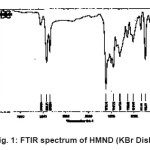
Figure 1: FTIR Spectrum of HMND (KBr Disk)
Figure 2: FTIR Spectrum of [UO2 (HMND)]2+ (KBr Disk)
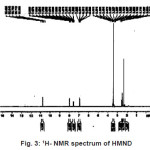
Figure 3: 1H- NMR Spectrum of HMND
Figure 4: 1H- NMR Spectrum of [UO2(HMND)]2+
Cell Culture
The human tumor cell line (H29: human colon adenocarcinoma cell line, T47D: human breast adenocarcinoma cell line, used for treatment with the drug was provided. H29 and T47D Cell were grown at 37 °C in an atmosphere containing 5% CO2, wet 95% with RPMI-1640 Medium HEPES Modification with 2mM L-glutamine and 25 mM HEPES (Sigma-Aldrich Chemie GmbH, Germany) supplemented with 10% heat-inactivated fetal bovine serum (FBS) (Gibco, Carlsbad, Calif, USA), 20gr/lit sodium bicarbonate, and 500 mg/L (100uint/ ml) ampicillin, ester petomysin 100micro gram/ml.
Cytotoxicity Studies
HMND ligand and [UO2 (HMND)]2+ complex are two compounds which were assayed for cytotoxicity in vitro against (HT29: Haman colon adenocarcinoma) cells and T47D: human breast adenocarcinoma cell line) cells. The two cell lines were provided by the Pasteur Institute in Iran. The procedure for cytotoxicity studies was similar to that reported earlier.15 Briefly, in order to calculate the concentration of each drug that produces a 50% inhibition of cell growth (IC50), 190 mL of cell suspension 4×10⁵ cell/cm3) was exposed to various concentrations of ligand and complexes dissolved in sterile DMSO. The final concentration of DMSO in the growth medium was 2% (v/v) or lower, concentrations without effect on cell replication. After the incubation period’s 72 hours for all cell lines, the cell concentrations were determined both in control and in drug-treated cultures. All experiments were done for six times.
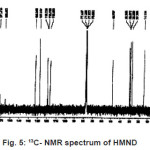
Figure 5: 13C- NMR Spectrum of HMND
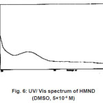
Figure 6: UV/ Vis Spectrum of HMND (DMSO, 5×10⁻⁴ M)
Figure 7: UV/ Vis Spectrum of [UO2(HMND)]2+ (DMSO, 5×10⁻⁴ M)
Figure 8: Thermal Analysis Data of [UO2(HMND)]2+
Synthesis of the [UO2 (HMND)]2+
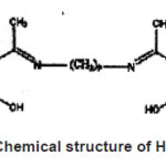
For synthesis of the [UO2 (HMND)]2+ to a magnetically stirred of ligand (0.63g, 1.3mmol) in acetonitrile(10ml) was added uranyl (VI) nitrate (0.5g, 1.3 mmol) at room temperature. The reaction mixture was further stirred for 3 hours to ensure the completion and precipitation of the formed complex. The precipitated solid complex was filtered and washed several times with diethyl ether to remove any traces of the unreacted starting materials.Yield, 88%. Anal. Calcd of [UO2 (HMND)]2+; C;40.20, H; 5.11, N; 2.1; found: C; 40.46, H. 5.68, N; 2.76. Mp: 210 °C. ¹HNMR (DMSO): 6.7-7.6 (CH phenol), 1.7 (CH3), 3.5 (N-CH2), 2.7 (CH2), FT-IR (KBr, cm-1): 1284s (νC-N), 1630 s (νC=N), 2928 br(νOH), 574 m (νU-O), 472 w (νU-N), 919 s (νO=U=O), UV-vis (DMSO): λmax 265nm (ε27000), 320nm(ε7000), 400nm(ε3400) (Fig. 1-9). [UO2 (HMND)]2+ is soluble in acetone, chloroform, ethanol, dichloro methane, diethyl ether, DMF and DMSO and insoluble in water, Acetonitrile and hexane and little soluble in methanol. Fig. 10, 11 shows Chemical structures of HMND and [UO2 (HMND)]2+.
Figure 9: Chemical Structure of HMND
Figure 10: Chemical Structure of [UO2(HMND)]2+
Figure 11: Morphology Cells (A: HT-29, B: T47-D) in Contain HMND (0/001M)
Analysis of HMND Ligand
Anal:%69. Calcd of C25H34N2O2; C; 76.16, H; 8.62, N; 3.55; found: C; 80.21, H. 9.30, N; 4.98. Mp83-85 °C, ¹HNMR (DMSO): 6.9-8 (CH phenol), 1.7 (CH3), 3.7(N-CH2), 2.5 (CH2),10.9 (OH), FT-IR (KBr, cm-1): 1307s (νC-N), 1612 s (νC=N), 2921 br(νOH), UV-vis(DMSO): λmax 260nm(ε15000), 313nm(ε7000). HMND is soluble in acetonitrile, DMF, DMSO, dichloro methane and chloroform and insoluble in acetone,water, methanol, ethanol, diethyl ether and hexane.
Results
Preparation of Ligand and Complex
The reaction of uranyl nitrate with the ligand in acetonitrile solvent result the formation of [UO2L] in that L=HMND in the molar ratio 1:1(metal: ligand). Compounds are quite stable and could be stored without any appreciable changes for long time. Compounds were characterized by several techniques using FT-IR, UV-Visible and NMR spectra Thermal analysis were studied for these compounds. The [UO2(HMND)]2+ has210°C melting points respectively. It is soluble in acetone, chloroform, ethanol, dichloro methane, diethyl ether, DMF and DMSO and insoluble in water, Acetonitrile and hexane and little soluble in methanol. The spectral data of the complexes have good relationship with the literature data.
Cytotoxicity Assays in Vitro
HMND and [UO2 (HMND)]2+ are compounds which were assayed for cytotoxicity in vitro against (HT29) cells and T47D) cells. The two cell lines were provided by the Pasteur Institute in Iran. The procedure for cytotoxicity studies was similar to that reported earlier. The concentration ranges of 0.01, 0.001 M for HMND and complex in DMSO were prepared. After preincubation lasting for 12 (24) hours at 37 °C in 5% CO2 atmosphere and 95% humidity the tested compounds. The color intensity was measured by Eliza instrument in 570 and 630 nm. The mechanism by which this complex acts as antitumor agents is apoptosis from the ligand. The life percentage of cell (HT-29) of HMND in 0.01 and 0.001 is 13.9 and 33.7 respectively. The life percentage of cell (742) of HMND in 0.01 and 0.001 is 14.4 and 18.4 respectively. The life percentage of cell (HT-29) of [UO2 (HMND)]2+in 0.01 and 0.001 is 19.5 and 14.8 respectively. The life percentage of cell (742) of [UO2 (HMND)]2+ in 0.01 and 0.001 is 14.5 and 11 respectively. (Fig. 12).
Thermo Gravimetric Analyses
The thermal properties of these compounds were investigated by thermo grams (TG, DTG andDTA). Fig. 9. shows TGA and DTA curves for[UO2 (HMND)]2+. In the temperature range 200-260°C, 10% weight losing was observed which was related to the loss ofmost parts of compound. In the temperature range from 300-440°C, 67.3% weight reduction was found, which was related to
the loss of a part of compound.
Discussion
In this research, some of the inorganic complexes of uranyl with N- donor ligands were synthesized. Complexes were characterized by FTIR and UV, ¹HNMR, ¹³CNMR spectra, TG/DTG measurements and some physical properties. The results of simultaneous TG-DTG-DTA analyses of the complexes show the final degradation product for these complexes are UO3. The antitumor activity of used ligands and their complexes against a panel of human tumor cell lines (HT29: Haman colon adenocarcinoma cell line T47D: human breast adenocarcinoma cell line) were studied and determined by MTT (3-[4,5-dimethylthiazol-2-yl]-2,5- diphenyl-tetrazolium bromide) assay.
Acknowledgments
We gratefully acknowledge the financial support from the Research Council of malard Islamic Azad University and many technical supports that provided by Tarbiat Modarres University.
References
- Vidmar R. J. IEEE Trans. Plasma Sci., 1992; 21: 876 (1992).
- Murthy A. S. N. and Reddy A. R. Journal of Chemical Sciences., 1981; 90: 519 (1981).
- Razakantoanina V. N. K. and Phung P. Parasitology Research., 2000; 86: 665 (2000).
- Royer R. E. and Deck L. M. Journal of Medicinal Chemistry., 1995; 38: 2427 (1995).
- Flack M. R. and Pyle R. G. The Journal of Clinical Endocrinology & Metabolism., 1995; 76: 1019 (1995).
- Baumgrass R. and Weiwad M. Journal of Biological Chemistry., 2001; 276: 47914 (2001).
- Teimouri M. B. and Bazhrang R. Bioorganic Medicinal Chemistry Letters., 2006; 16: 3697 (2006).
- Teimouri M. B. Tetrahedron., 2006; 62: 10849 (2006).
- Grevy J. M., Tellez F. Inorganica Chimica Acta., 2002; 339: 532 (2002).
- Bernalte A. and Barros F. J. Polyhedron., 1999; 18: 2907 (1999).
- Lemma K. and Berglund J. Journal of Biological Inorganic Chemistry., 2000; 5: 300 (2000).
- Campbell M. J. M. Coordination Chemistry Reviews., 1975; 15: 279 (1975).
- Padhy´e S. and Kauffman G. B. Coordination Chemistry Reviews., 1985; 63: 127 (1985).
- Erwin B. and Omoshile C. Journal of the Chemical Society Perkin Transactions., 1995; 2: 1333 (1995).
- Zhao G. and Lin H. Journal of Inorganic Biochemistry., 1998; 70: 219 (1998).

This work is licensed under a Creative Commons Attribution 4.0 International License.

![Fig. 2: FTIR spectrum of [UO2 (HMND)]2+ (KBr Disk)](http://www.materialsciencejournal.org/wp-content/uploads/2012/12/Vol13_No1_Syn_SHA_fig2-150x150.jpg)
![Fig. 4: 1H- NMR spectrum of [UO2(HMND)]2+](http://www.materialsciencejournal.org/wp-content/uploads/2012/12/Vol13_No1_Syn_SHA_fig4-150x150.jpg)
![Fig. 7: UV/ Vis spectrum of [UO2(HMND)]2+ (DMSO, 5×10⁻⁴ M)](http://www.materialsciencejournal.org/wp-content/uploads/2012/12/Vol13_No1_Syn_SHA_fig7-150x150.jpg)
![Fig. 8: Thermal analysis data of [UO2(HMND)]2+](http://www.materialsciencejournal.org/wp-content/uploads/2012/12/Vol13_No1_Syn_SHA_fig8-150x150.jpg)
![Fig. 10: Chemical structure of [UO2(HMND)]2+](http://www.materialsciencejournal.org/wp-content/uploads/2012/12/Vol13_No1_Syn_SHA_fig10-150x150.jpg)

