Komal Grover1, and Kiran Jeet2*
1Department of Mathematics, Statistics, and Physics, Punjab Agricultural University, Ludhiana, Punjab, India.
2Electron Microscopy and Nanoscience Laboratory, Punjab Agricultural University, Ludhiana, Punjab, India.
Corresponding Author Email: kiranjeet@pau.edu
DOI : http://dx.doi.org/10.13005/msri/200107
Article Publishing History
Article Received on : 24 Jan 2023
Article Accepted on : 14 Mar 2023
Article Published : 10 Apr 2023
Plagiarism Check: Yes
Reviewed by: Dr. Prashant Koli
Second Review by: Dr. Nadiah Bte Ameram
Final Approval by: Dr. K. M. Garadkar
Article Metrics
ABSTRACT:
Adsorption is considered one of the best methods for the removal of heavy metal ions from an aqueous solution. However, the synthesis of adsorbents with desired selectivity and performance remains a key challenge in the battle of water decontamination. Recently, carbon-based and metal-oxide based nanomaterials have emerged as promising candidates for the adsorption of heavy metals due to their high specific surface area, high aspect ratio, and concentrated pore size distribution. Here, in this work five adsorbents ie. Graphene Oxide (GO), Magnetic Graphene Oxide (MGO), Titanium Dioxide (TiO
2), and their composites GO-TiO
2 and MGO-TiO
2 were synthesized. The prepared samples were characterized via high-resolution imaging, BET-N
2 adsorption-desorption analysis, and spectroscopic techniques. TEM results revealed the nanoscale structures of the synthesized nanomaterials. The approximate sizes of MGO and TiO
2 nanoparticles found under TEM studies were about 24.58 and 35.51 nm respectively. The presence of desired functional groups was very well deciphered by FT-IR spectroscopy. Results of N
2 adsorption-desorption studies revealed that the prepared GO was macro-porous while all other samples were mesoporous. MGO was found to have the highest BET surface area of about 108.375 m
2/g. These results indicate that the prepared nanomaterials may serve the purpose of effectively adsorbing the heavy metal ions from an aqueous solution.
KEYWORDS:
Adsorption; Graphene Oxide; Heavy metals; Nanomaterials; Titanium Dioxide
Copy the following to cite this article:
Grover K, Jeet K. Preparation and Characterization of Nanohybrids Made of Graphene Oxide as Super Adsorbents. Mat. Sci. Res. India; 20(1).
|
Copy the following to cite this URL:
Grover K, Jeet K. Preparation and Characterization of Nanohybrids Made of Graphene Oxide as Super Adsorbents. Mat. Sci. Res. India; 20(1). Available from: https://bit.ly/3KxneJZ
|
Introduction
The discharge of heavy metals into aquatic ecosystems has raised global concerns over the past few decades. These contaminants are introduced into water bodies through effluents from various industries, including paper and pulp, petrochemicals, automobiles, and battery manufacturing. While several methods, such as precipitation, ion exchange, reverse osmosis, and membrane filtration, have been employed to remove heavy metals. Most of these methods have drawbacks, such as high cost, low efficiency, and sludge generation (Kumar et al., 2019). Additionally, they fail to meet the demand for water resources, particularly for large volumes. Adsorption, on the other hand, is a simple, economical, and adaptable method in terms of unit design. Natural and synthetic adsorbents, such as clay, activated carbon, mesoporous silica, and resin, have been used to treat heavy metal-contaminated water. However, separating and regenerating these adsorbents from wastewater poses a significant challenge. Therefore, there is a need to develop novel adsorbents with high adsorption capacity and quick separation from large volumes of water (Almomani et al., 2019). Recently, nanomaterials, particularly carbon-based and metal oxide-based ones, have emerged as promising adsorbents for heavy metal ions due to their high specific surface area, surface-to-volume ratio, and concentrated pore size distribution (Khan et al., 2013; Qu et al., 2015). Among these, graphene oxide (GO), a carbon-based nanomaterial, has received widespread attention (Yan et al., 2014, Majumder P, Gangopadhyay R. 2022). GO consists of a hexagonal network of covalently bonded carbon atoms with oxygen-containing functional groups, such as hydroxyl, epoxy, lactone, quinone, phenol, anhydride, carbonyl, ether, and carboxyl groups, attached to various sites, which facilitate the binding of positively charged metal ions to its surface (Guerrero-Fajardo et al., 2020; Zhao et al., 2011). However, GO’s hydrophilicity and tendency to agglomerate during its application and storage make it challenging to separate, even after saturation adsorption (Liu et al., 2015; Sun et al., 2015). To address these issues, GO can be magnetized, for example, with iron oxides. Nano-sized iron oxides exhibit super-paramagnetism, low toxicity, chemical inertness, and the ability to immobilize various adsorbents on their surface (Jawed et al., 2020). The magnetized GO can be easily separated using an external magnetic field (Lingamdinne et al., 2019). Another category of nanomaterials that has shown favourable adsorption towards heavy metals is nano-sized metal oxides (Hua et al., 2012). Functionalizing GO with metal oxides largely increases the electronegative charge on its surface thereby improving metal removal efficiency (Jawed et al., 2020). The inclusion of active materials such as manganese dioxide (Xiang et al., 2018), iron oxide (Tian et al., 2017), and titanium dioxide (Liu et al., 2016) have sparked a profound interest in the field of adsorption as the metallic compounds could improve the adsorption performance by providing more active sites (Lai et al., 2020). Titanium dioxide nanoparticles are widely used as an adsorbent for heavy metal ions due to their low cost, stability, and non-toxicity towards human beings and the environment (Seidlerová et al., 2016). Here in this work the synthesis of adsorbents such as Graphene Oxide (GO), Magnetic Graphene Oxide (MGO), Titanium Dioxide (TiO2), and their composites GO-TiO2, and MGO-TiO2 has been reported. GO-TiO2 is synthesized keeping in view the favourable properties of both GO and TiO2. MGO-TiO2 nanocomposites are synthesized aiming at integrating the advantages of all the components – GO, TiO2 along with magnetic properties of iron oxide for the added advantage of easier regeneration of adsorbents and improve the overall adsorption efficiency.
Material and Methods
Graphite fine powder extra pure of size 10-30 nm was procured from Loba Chemicals, sulfuric acid (H2SO4), potassium permanganate (KMnO4), phosphoric acid (H3PO4), ferrous ammonium sulfate hexahydrate [(NH4)2Fe(SO4)2.6H2O], ammonium ferric sulfate dodecahydrate [(NH4 Fe(SO4)2).10H2O], aqueous ammonia (NH4OH) (weight 25%), hydrogen peroxide (H2O2), isopropyl alcohol (IPA), titanium isopropoxide (TTIP), isopropanol, HCl, and anhydrous ethanol were procured from Molychem, and were of analytical grade and used without any further purification.
Synthesis of Adsorbents
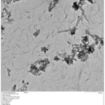
GO was prepared by the modified Hummer’s method. Graphite powder was added into a mixture of concentrated H2SO4 and H3PO4 in the ratio of 9:1 under continuous stirring followed by the addition of KMnO4 to bring about the oxidation. The mixture was stirred continuously till the color changed to purple. Then the mixture was cooled down to room temperature and an ice water mixture and H2O2 were added to stop the reaction. A yellow-colored solution was obtained which was then sonicated to bring about the complete exfoliation of graphite oxide. Then the solution was centrifuged once with HCl and many times with distilled water until a neutral pH was obtained. Finally, solid GO was obtained by vacuum drying in an oven and grinding using a pestle and mortar (Kaur and Jeet 2017). The observations recorded during the synthesis of GO are given in figure 1.
MGO was prepared by the co-precipitation of iron oxide nanoparticles on the surface of GO. A dispersion of GO was prepared by adding a small amount of prepared GO in 100 ml of double-distilled water and sonicating it for an hour. Then, 10.7 g of ferric ammonium sulfate and 5.8 g of ferrous ammonium sulfate were added to 100 ml of double-distilled water followed by the rapid addition of 10 ml of aqueous ammonia. The solution was then kept under stirring and GO suspension was slowly added to this solution after that the stirring was continued for about 45 minutes at 85 0C and then the solution was kept undisturbed overnight. Finally, MGO was separated with a magnet and washed thrice with distilled water and anhydrous ethanol respectively. Subsequently, MGO was kept in an oven at 70 0C for drying (Deng et al., 2013, Yi et al. 2021). The observations recorded during the synthesis of MGO are given in figure 2.
Titanium Dioxide was prepared by a wet chemical method. 10 ml of TTIP was added into a solution containing ethanol and double distilled water in the ratio of 7:1 under constant stirring. The stirring was further continued at room temperature until a thick paste with lots of nanoparticles was obtained, which were then separated by centrifugation with distilled water. The nanoparticles were then calcined and stored in an airtight container (Tamilselvi et al., 2016, MironyukI et al. 2020). The observations recorded during the synthesis of TiO2 are given in figure 3.
GO-TiO2 nanocomposite was prepared by the sol-gel process. A small amount of the prepared GO was added into the double distilled water and was sonicated to re-exfoliate GO. Afterward, a mixture of isopropanol and TTIP in the ratio of 4:1 was dropped into the GO solution under continuous stirring for the crystallization of TiO2 Nanoparticles. The precipitates were washed, filtered with water and ethanol, and dried in the oven. Finally, GO-TiO2 nanohybrids were obtained by calcination at 4000C (Sakulpaisan et al., 2016). The observations recorded during the synthesis of GO-TiO2 are given in figure 4.
For the preparation of MGO-TiO2 nanocomposite, the prepared GO and TiO2 nanoparticles were separately dispersed in double-distilled water and were sonicated for about an hour. Then (NH4)2Fe(SO4)2.6H2O and (NH4Fe(SO4)2).10H2O were dispersed in distilled water and 10 ml of aqueous ammonia was quickly added to this solution, as in the preparation of MGO. Afterward, this solution and TiO2 dispersion were simultaneously added to the GO dispersion under vigorous stirring. The stirring was further continued at 850C for 45 minutes and kept overnight at room temperature. The rest of the procedure of obtaining nanoparticles was the same as that in the case of MGO (Chang et al., 2015). The pictorial representation of MGO-TiO2 synthesis is given in figure 5.
Characterization
The surface area of the prepared nanomaterials was examined by BET N2 adsorption-desorption analysis using Micromeritics ASAP 2020 volumetric adsorption analyzer installed at Emerging Life Sciences, Guru Nanak Dev University, Amritsar, Punjab, India. Before each analysis, each sample was degassed for 4-6 hours at 2000C. For the determination of the elemental composition and functional groups present the prepared samples were analyzed using Perkin Elmer Fourier Transform Infrared Spectrometer installed at Central Instrumentation Facility, Lovely Professional University, Phagwara, Punjab, India. The prepared samples were viewed under Transmission Electron Microscopy (TEM) installed at the Electron Microscopy and Nanoscience Lab, Punjab Agricultural University, Ludhiana, Punjab, India, to study their structural and morphological properties.
Results and Discussions
BET N2 Adsorption-Desorption Analysis
Magnetic Graphene Oxide was found to have the largest surface area of 108.3750 m2/g. The magnetization of GO appeared to increase its surface area, as evidenced by the larger surface area of MGO compared to GO. The surface areas of both the nanocomposites ie. GO-TiO2 and MGO-TiO2 were less than that of MGO which showed that the substitution of graphene oxide with titanium dioxide decrease its surface area. Due to the agglomeration of individual nanoparticles, the surface area of TiO2 nanoparticles was only 4.42 m2/g. The extremely high calcination temperature may have contributed to the high average particle size of TiO2 particles. As a result, the individual TiO2 nanoparticles were agglomerated which was also evident from the TEM micrograph of TiO2 nanoparticles. The pore diameters of the synthesized samples confirmed the presence of mesopores as per the IUPAC classification except for GO which was found to be macroporous (Thommes et al., 2015). The BET surface areas, pore sizes, and pore volume distribution of the synthesized samples are given in Table 1.
Table 1: BET surface areas, pore sizes, and pore volume distribution of the synthesized samples.
|
Adsorbent
|
BET surface area (m2/g)
|
Pore width (nm)
By BET method
|
Pore volume (cm3/g) By BET method
|
Average particle size (nm)
|
BJH adsorption average pore diameter (nm)
|
|
Graphene Oxide (GO)
|
35.8220
|
31.97
|
0.286316
|
167.4947
|
51.7066
|
|
Magnetic Graphene Oxide (MGO)
|
108.3750
|
9.77
|
0.264600
|
55.36
|
9.67
|
|
Titanium Dioxide (TiO2)
|
4.4288
|
268.67
|
0.297472
|
1354.76
|
46.755
|
|
GO-TiO2
|
106.1106
|
16.89
|
0.448001
|
56.545
|
13.91
|
|
MGO-TiO2
|
81.3364
|
19.71251
|
0.400836
|
73.77
|
23.63
|
Transmission Electron Microscopy
Figure 6 shows the TEM micrographs of the prepared samples. The TEM micrograph of GO captured at 500 nanometers (Figure 6a) reveals a layered-wrinkled structure with many folds on it, which may be attributed to various oxygen-containing functional groups (Liu et al., 2015; Deng et al., 2013, Nhlane et al. 2021 ). The TEM micrograph of MGO (Figure 6b) was captured at 100 nm and depicts the homogeneous distribution of iron-oxide nanoparticles on the surface of GO. The average diameter of MGO nanoparticles determined using Image J software was found to be 24.58 nm (Deng et al., 2013). The TEM micrograph of TiO2 nanoparticles (Figure 6c) reveals that the formed TiO2 nanoparticles are almost spherical and are aggregated to some extent. The average diameter of TiO2 nanoparticles was found to be 35.51 nm. The TEM micrograph of GO-TiO2 nanocomposite (Figure 6d) shows the successful deposition of TiO2 nanoparticles on the layered structure of GO (Ibrayev et al., 2019). The TEM images of the MGO-TiO2 nanohybrid (Figure 6e) depict the presence of iron-oxide nanoparticles, some iron rods, and TiO2 nanoparticles on the surface of GO, showing the successful stacking of all the components. The crumpled structure of GO sheets enables the binding of nanoparticles onto their surface (Chang et al., 2015; Jo & Selvam, 2015).
FT-IR spectroscopy
The FT-IR spectra of the prepared samples are presented in Figure 7. The FT-IR spectrum of GO displayed a broad band at 3225.37 cm-1, which can be attributed to the presence of water adsorbed on the surface of GO or to the structural hydroxyl groups (-COH and -COOH) of GO. Additionally, a band at 1722.77 cm-1 was observed, which is characteristic of the vibrations of the C=O group, and a band at 1046.22 cm-1 was due to C-O stretching vibrations of the alkoxy group (Sitko et al., 2013).
The FT-IR spectrum of MGO exhibited a band at 3203.39 cm-1, which can be attributed to the O-H stretching vibrations. Additionally, a band at 551.62 cm-1 was observed, which is due to Fe-O stretching vibrations. The band at 890.95 cm-1 supports the deposition of iron nanoparticles on the surface of GO (Liu et al., 2015; Sun et al., 2018).
The FT-IR spectrum of TiO2 exhibited only one major peak at 499 cm-1, which is characteristic of Ti-O-Ti bonds (Bok-Badura et al., 2017). The FT-IR spectrum of GO-TiO2 showed bands at 1635.26 cm-1 and 3387 cm-1, which are characteristic of aromatic groups and O-H stretching vibrations, respectively. Additionally, a band at 523.51 cm-1 was observed, which corresponds to the Ti-O-Ti vibrations (Sakulpaisan et al., 2016; Kurniawan et al., 2020).
The FT-IR spectrum of MGO-TiO2 displayed bands at 3058 cm-1, 1578 cm-1, 1086 cm-1, and 788 cm-1, which are attributed to the adsorbed water content on the GO surface, C=C vibrations, C-O vibrations, and C-O-Ti vibrations, respectively. The band at 884 cm-1 can be attributed to the covalent bonding of Fe3O4 nanoparticles on the GO surface. The lowering of the carbonyl band of GO to 1431 cm-1 indicates the successful deposition of TiO2 and Fe3O4 nanoparticles on the GO surface (Zhang et al., 2015).
Conclusion
The morphologies and size of the prepared nanomaterials were determined using transmission electron microscopy. TEM micrograph of GO depicted the formation of the layered structure of GO with many folds which might be attributed to the presence of oxygen-containing functionalities, TiO2 micrograph depicted the formation of large agglomerates of TiO2 particles. MGO-TiO2 revealed the deposition of iron oxide nanoparticles and TiO2 nanoparticles on the surface of GO. From the TEM micrographs the approximate sizes of MGO and TiO2 nanoparticles were found to be about 24.58 and 35.51 nm respectively. FT-IR spectra of the synthesized nanomaterials depict the presence of required functional groups on their respective surfaces. N2 adsorption-desorption studies exhibited that MGO was having the highest surface area (108.3750 m2/g), and the prepared GO was macroporous while all other samples were mesoporous. Thus, the successful preparation of the adsorbents was confirmed.
Acknowledgments
We sincerely thank the Department of Science and Technology for providing us research grant under the Promotion of University Research and Scientific Excellence (PURSE) grant scheme for carrying out the research work of this manuscript.
Conflict of interest
On behalf of all authors, the corresponding author states that there is no conflict of interest.
Funding Sources
Department of Science and Technology (DST) under research grant Promotion of University Research and Scientific Excellence (PURSE).
References
- Almomani F, Bhosale R, Khraisheh M, kumar A, Almomani T (2019) Heavy Metal ions removal from industrial wastewater using magnetic nanoparticles (MNP), Appl. Surf. Sci, doi: https://doi.org/10.1016/ j.apsusc.2019.144924
CrossRef - Argun M E and Dursun S (2008) A new approach to modification of natural adsorbent for heavy metal adsorption. Bioresour in Technol 99 2516-2527 https://doi.org/10.1016/j.biortech.2007.04.037.
CrossRef - Bok-Badura J, Jakóbik-Kolon A, Karoń K and Mitko K (2017) Sorption studies of heavy metal ions on pectin-nano-titanium dioxide composite adsorbent in Sep Sci Technol 53 1034-1044 http://dx.doi.org/10.1080/01496395.2017.1329840.
CrossRef - Chang Y N, Ou X M, Zeng G M, Gong J L, Deng C H, Jiang Y, Liang J, Yu G Q, Liu H Y and He X (2015) Synthesis of magnetic graphene oxide–TiO2 and their antibacterial properties under solar irradiation in Appl Surf Sci 343 1-10 https://doi.org/10.1016/j.apsusc.2015.03.082.
CrossRef - Deng J H, Zhang X R, Zeng G M, Gong J L, Niu Q Y and Liang J (2013) Simultaneous removal of Cd(II) and ionic dyes from aqueous solution using magnetic graphene oxide nanocomposite as an adsorbent in J Chem Eng 226 189-200 https://doi.org/10.1016/j.cej.2013.04.045.
CrossRef - Guerrero-Fajardo C A, Giraldo L and Moreno-Piraján J C (2020) Preparation and Characterization of Graphene Oxide for Pb(II) and Zn(II) Ions Adsorption from Aqueous Solution: Experimental, Thermodynamic and Kinetic Study in Nanomaterials 10(6) 1022 https://doi.org/10.3390/nano10061022.
CrossRef - Hua M, Zhang S, Pan B, Zhang W, Lv L and Zhang Q (2012) Heavy metal removal from water/wastewater by nanosized metal oxides: A review in J Hazard Mater 211–212 317–331 https://doi.org/10.1016/j.jhazmat.2011.10.016.
CrossRef - Ibrayev N, Zhumabekov A, Ghyngazov S and Lysenko E (2019) Synthesis and study of the properties of nanocomposite materials TiO2-GO and TiO2-rGO. Mater Res Express, 6 1-10 https://doi.org/10.1088/2053-1591/ab51a3.
CrossRef - Jawed A, Saxena V and Pandey L M (2020) Engineered nanomaterials and their surface functionalization for the removal of heavy metals: A review in J Water Process Eng 33 101009 https://doi.org/10.1016/j.jwpe.2019.101009.
CrossRef - Jo W K and Selvam N C S (2015) Synthesis of GO supported Fe2O3–TiO2 nanocomposites for enhanced visible-light photocatalytic applications in Dalton Trans 44 16024-16035 https://doi.org/10.1039/C5DT02983J.
CrossRef - Kaur K and Jeet K (2017) Electrical conductivity of water- based nanofluids prepared with graphene – carbon nanotube hybrid. Fuller Nanotub in Carbon Nanostructures 2 726-734 https://doi.org/10.1080/1536383X.2017.1389906.
CrossRef - Khan M A, Gee E, Choi J, Kumar M, Jung W, Timmes T C, Kim H-C, & Jeon B-H (2013) Adsorption of cobalt onto graphite nanocarbon-impregnated alginate beads: equilibrium, kinetics, and thermodynamic studies in Chem Eng. Commun 201 (3) 403–418 https://doi.org/10.1080/00986445.2013.773426.
CrossRef - Kumar M, Chung J S and Hur S H (2019) Graphene Composites for Lead Ions Removal from Aqueous Solutions in Appl Sci 9(14) 1-30 https://doi.org/10.3390/app9142925.
CrossRef - Kurniawan T A, Mengting Z, Fu D, Yeap S K, Othman M H D, Avtar R and Ouyang T (2020) Functionalizing TiO2 with graphene oxide for enhancing photocatalytic degradation of methylene blue (MB) in contaminated wastewater in J Environ Manage 270 110871 https://doi.org/10.1016/j.jenvman.2020.110871.
CrossRef - Lai K C, Lee L Y, Hiew B Y Z, Thangalazhy-Gopakumar S, Gan S (2020) Facile synthesis of xanthan biopolymer integrated 3D hierarchical graphene oxide/titanium dioxide composite for adsorptive lead removal in wastewater in Bioresource Technology 309 123296. https://doi.org/10.1016/j.biortech.2020.123296.
CrossRef - Lingamdinne L P, Koduru J R and Karri R R (2019) A comprehensive review of applications of magnetic graphene oxide based nanocomposites for sustainable water purification in J Environ Manage 231 622-634 https://doi.org/10.1016/j.jenvman.2018.10.063.
CrossRef - Liu J, Ge X, Ye X, Wang G, Zhang H, Zhou H, Zhang Y and Zhao H (2016) 3D graphene/δ-MnO2 aerogels for highly efficient and reversible removal of heavy metal ions in J Mater Chem A 4 1970-1979 https://doi.org/10.1039/C5TA08106H.
CrossRef - Liu L, Zhang Y, He Y, Xie Y, Huang L, Tan S and Cai X (2015) Preparation of montmorillonite-pillared graphene oxide with increased single- and co-adsorption towards lead ions and methylene blue in RSC Adv 5 3965-3973 https://doi.org/10.1039/C4RA13008A.
CrossRef - Majumder P, Gangopadhyay R (2022) Evolution of graphene oxide (GO)-based nanohybrid materials with diverse compositions: an overview. RSC Adv 12:5686–5719 https://doi.org/10.1039/D1RA06731A
CrossRef - MironyukI. F., SoltysL. M., TatarchukT. R., & SavkaK. O. (2020). Methods of Titanium Dioxide Synthesis (Review). Physics and Chemistry of Solid State, 21(3), 462-477. https://doi.org/10.15330/pcss.21.3.462-477
CrossRef - Nhlane D, Richards H, Etale A (2021) Facile and green synthesis of reduced graphene oxide for remediation of Hg(II)-contaminated water. Mater Today Proc 38:737–742 https://doi.org/10.1016/j.matpr.2020.04.163
CrossRef - Qu J, Zhang Q, Xia Y, Cong Q and Luo C (2015) Synthesis of carbon nanospheres using fallen willow leaves and adsorption of Rhodamine B and heavy metals by them in Environ Sci Pollut Res Int 22 1408–1419 https://doi.org/10.1007/s11356-014-3447-x.
CrossRef - Roy A and Bhattacharya J (2015) Nanotechnology in industrial wastewater treatment. IWA Publishing, London, UK
CrossRef - Sakulpaisan S, Vongsetskul T, Reamouppaturm S, Luangkachao L, Tantirungrotechai J and Tangboriboonrat P (2016) Titania-functionalized graphene oxide for an efficient adsorptive removal of phosphate ions in J Environmental Management 167 99–104. https://doi.org/10.1016/j.jenvman.2015.11.028.
CrossRef - Seidlerová J, Šafařík I, Rozumová L, Šafaříková M and Motyka O (2016) TiO2-Based Sorbent of Lead Ions in Procedia Mater Sci 12 147 – 152 https://doi.org/10.1016/j.mspro.2016.03.026.
CrossRef - Sitko R, Turek E, Zawisza B, Malicka E, Talik E, Heimann J, Gagor A, Feist B and Wrzalik R, (2013) Adsorption of divalent metal ions from aqueous solutions using graphene oxide in Dalton Trans 42 5682-5689 https://doi.org/10.1039/C3DT33097D.
CrossRef - Sun J, Liang Q, Han Q, Zhang X and Ding M (2015) One-step synthesis of magnetic graphene oxide nanocomposite and its application in magnetic solid phase extraction of heavy metal ions from biological samples in Talanta 132 557-563 https://doi.org/10.1016/j.talanta.2014.09.043.
CrossRef - Sun M, Li P, Jin X , Ju X, Yan W, Yuan J and Xing C (2018) Heavy metal adsorption onto graphene oxide, amino group on magnetic nanoadsorbents and application for detection of Pb(II) by strip sensor in Food Agric Immunol 29 1053-1073 https://doi.org/10.1080/09540105.2018.1509946.
CrossRef - Tamilselvi S, Asaithambi M, and Sivakumar P (2016) Nano-TiO2-loaded activated carbon fiber composite for photodegradation of a textile dye in Desalination water treat 57 (33) 15495-15502 https://doi.org/10.1080/19443994.2015.1071684.
CrossRef - Thommes M, Kaneko K, Neimark A, Olivier J, Rodriguez-Reinoso F, Rouquerol J and Sing K (2015) Physisorption of gases, with special reference to the evaluation of surface area and pore size distribution (IUPAC Technical Report) in Pure Appl Chem 87 (9-10) 1051-1069 https://doi.org/10.1515/pac-2014–1117.
CrossRef - Tian C, Zhao J, Zhang J, Chu S, Dang Z, Lin Z and Xing B (2017) Enhanced removal of roxarsone by Fe3O4@3D graphene nanocomposites: synergistic adsorption and mechanism in Environ Sci: Nano 4 (11) 2134–2143 https://doi.org/10.1039/C7EN00758B.
CrossRef - Xiang C, Guo R, Lan J, Jiang S, Wang C, Du Z and Cheng C (2018) Self-assembling porous 3D titanium dioxide-reduced graphene oxide aerogel for the tunable absorption of oleic acid and RhodamineB dye in J Alloys Compd 735 246–252 http://dx.doi.org/10.1016/j.jallcom.2017.11.034.
CrossRef - Yan H, Li H, Tao X, Li K, Yang H, Li A, Xiao S, and Cheng R (2014) Rapid Removal and Separation of Iron(II) and Manganese(II) from Micropolluted Water Using Magnetic Graphene Oxide in ACS Appl. Mater. Interfaces 6 (12) 9871–9880 https://doi.org/10.1021/am502377n.
CrossRef - Yi He, Chen Yi, Xiliu Zhang, Wei Zhao, Dongsheng Yu (2021), Magnetic graphene oxide: Synthesis approaches, physicochemical characteristics, and biomedical applications, TrAC Trends in Analytical Chemistry,Volume 136,116191, https://doi.org/10.1016/j.trac.2021.116191
CrossRef - Zhang Y, Zhong C, Zhang Q, Chen B, He M and Hu B (2015) Graphene oxide–TiO2 composite as a novel adsorbent for the preconcentration of heavy metals and rare earth elements in environmental samples followed by on-line inductively coupled plasma optical emission spectrometry detection in RSC Adv 5 5996-6005 https://doi.org/10.1039/C4RA13333A.
CrossRef - Zhao G, Ren X, Gao X, Tan X, Li J, Chen C, Huang Y, and Wang X (2011) Removal of Pb(II) ions from aqueous solutions on few-layered graphene oxide nanosheets in Dalton Trans 40(41) 10945-10952 https://doi.org/10.1039/C1DT11005E.
CrossRef

This work is licensed under a Creative Commons Attribution 4.0 International License.